Forschungspublikationen

3 Ergebnisse
16.07.2026

OsteoMe: Multilayered GBR Membranes with Mucoadhesive and Osteoinductive Properties for Bone Augmentation

Vliesstoffe Composites Medizin

Zusammenfassung

The OsteoMe project, funded as part of the Joint Industrial Research (IGF) program, focuses on the development of a multilayer membrane that combines mucoadhesive, cell-excluding, and space-maintaining properties through a material- and structurally graded layered structure. With this property profile, combined with the use of bioresorbable materials, the project aims to meet all requirements for guided bone regeneration (GBR) membranes in dentistry and to specifically improve upon or surpass existing solutions. For manufacturing, electrospinning was combined with 3D printing and surface functionalization. As a result, three membrane layers were developed that individually represent specific aspects of the property profile and can be flexibly combined with one another.

Bericht

Introduction: The causes of tooth loss are very diverse, including trauma, tumor removal, tooth decay, and—very commonly—periodontitis (bacterial inflammation of the gums and jawbone). In Germany, approximately 50–60% of the population suffers from moderate to severe periodontitis, with the incidence increasing with age; consequently, a growing demand for artificial teeth is expected in the future [1]. In addition to the root cavity resulting from tooth loss, the causes mentioned above can also lead to damage to larger areas of the jawbone or necessitate their removal. Furthermore, bone resorption occurs due to the lack of mechanical stress on the jaw, making it impossible to place an implant without prior reconstruction of the bone tissue. Since an implant must be firmly anchored in the jawbone, prior bone augmentation is therefore absolutely necessary in these cases to provide patients with optimal care. In Germany, over 50% of dental implants placed require such augmentation [2–4]. In this treatment approach, a bone substitute material is filled into the cavity and covered with a GBR membrane. The membrane is essential for the success of the treatment, as its primary function is to prevent rapidly proliferating soft tissue cells from invading the cavity. Currently, both non-resorbable (e.g., PTFE, titanium) and resorbable (e.g., collagen) GBR membranes are used; however, they can only partially meet the required performance criteria. Resorbable membranes tend to be preferred because they do not need to be removed in a second surgical procedure—which could jeopardize treatment success—and carry a lower risk of infection. However, due to their lack of primary stability, their mechanical properties do not yet allow them to maintain space in a moist environment. For this reason, non-resorbable GBR membranes continue to be used as well [5, 6]. A GBR membrane that combines resorbability with space-maintaining properties in a way that meets clinical requirements does not yet exist, a fact underscored by high revision rates of approximately 20% [7, 8].

The IGF OsteoMe project aimed to develop novel, multilayer GBR membranes for bone augmentation with a complex, application-oriented property profile. To this end, the project partners ITM and FILK combined their complementary expertise in the development of electrospun and additive-manufactured (Fiber Additive Manufacturing, FAM) biopolymer structures, the functionalization of chitosans, and cell biological characterization. The property profile, which had been developed in advance through discussions with clinicians and industry representatives, was to be realized through a three-layer membrane structure consisting of a mucoadhesive layer for positional stabilization, a cell-excluding barrier layer, and a structuring, mineralized layer with defined porosity and mineral gradients to biomimetically replicate the bone-tissue interface and provide space-maintaining properties. Based on the materials polycaprolactone (PCL), silk fibroin (SF), and chitosan (Ch), simulation-based fabrication strategies were investigated that enable the targeted adjustment of mechanical, functional, and biological properties.

Results:

Mucoadhesive layer: Mucoadhesive materials allow for prolonged retention on mucous membranes and are therefore of interest for numerous medical applications. Chitosan is considered a promising mucoadhesive biopolymer due to its positive charge and the resulting interactions with negatively charged mucins. To further improve adhesion, the covalent binding of thiol-containing compounds was investigated, as these additionally enable the formation of disulfide bonds with mucin.

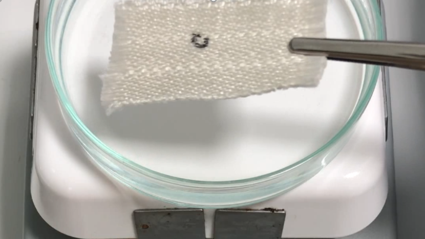
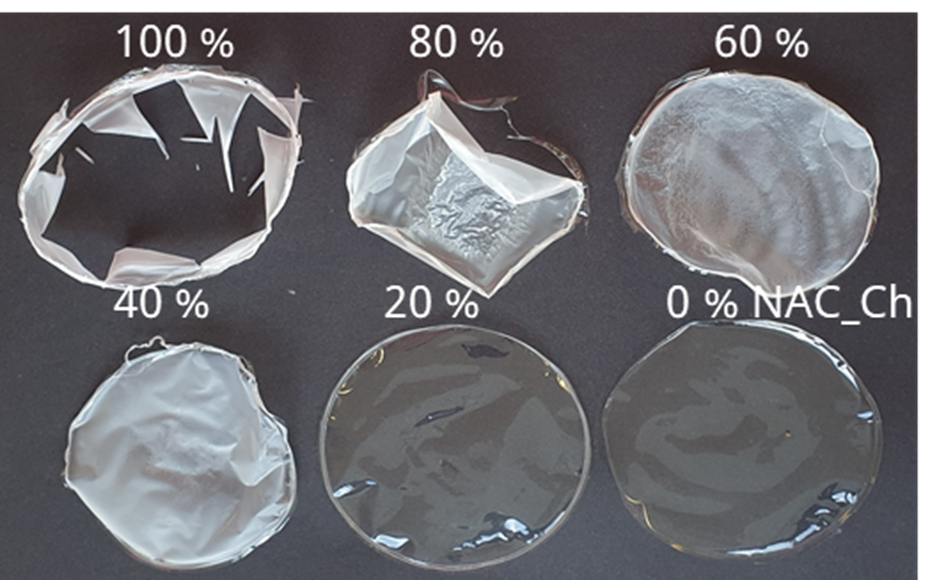
Functionalization was carried out via carbodiimide (EDC/NHS)-mediated coupling of N-acetylcysteine (NAC) or cysteine to chitosan with a degree of deacetylation exceeding 90%. Both the direct modification of chitosan films and the derivatization of dissolved chitosan were investigated. While the functionalization of films was complicated by the limited accessibility of the amino groups within the solid polymer matrix and resulted in an inhomogeneous distribution of thiol groups, the modification of dissolved chitosan enabled the preparation of reproducible conjugates with high thiol contents. The optimized chitosan conjugate achieved a total thiol content of approximately 878 µmol/g, with about 311 µmol/g present as free thiol groups. Due to the high reactivity of the thiol groups, a loss of approximately 20% was observed within two weeks during storage in air; therefore, storage under oxygen-free conditions is recommended. To produce mechanically stable membranes, the addition of at least 70 wt% unmodified chitosan was required (see Figure 1).

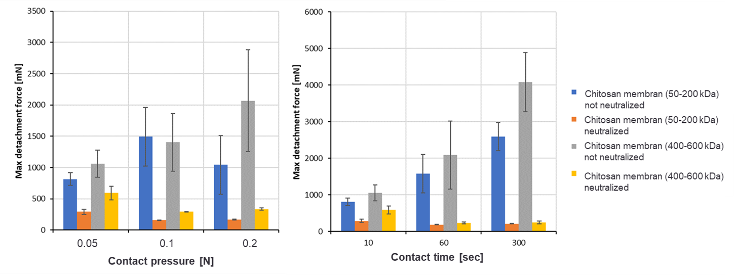
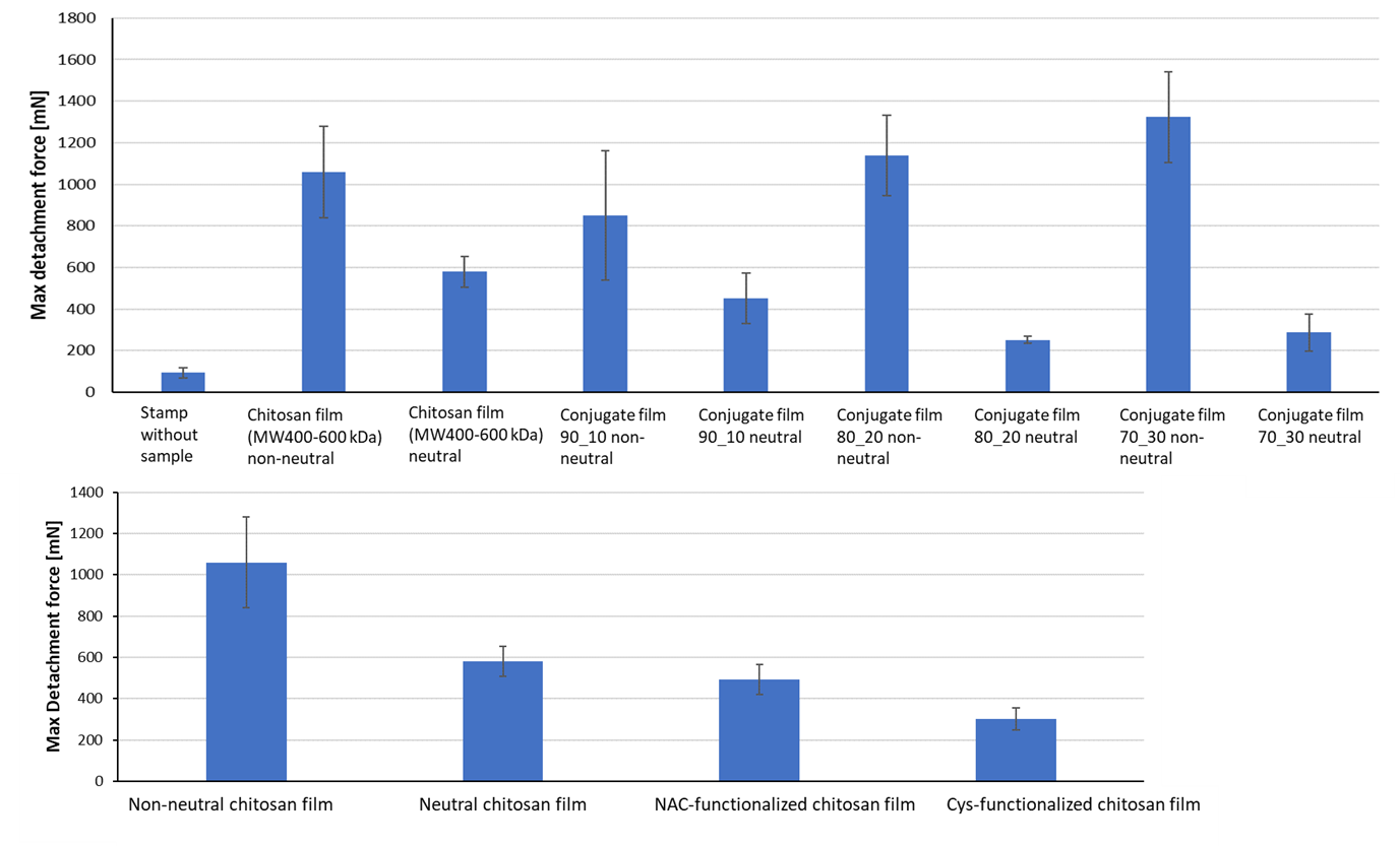
The mucoadhesive properties were investigated using a specially developed test setup on a texture analyzer, with porcine intestinal mucosa serving as the model substrate. The adhesive forces were determined relative to unmodified chitosan. In particular, acid-treated chitosan films exhibited high adhesion forces, which could be specifically influenced by varying the contact pressure and contact time. Neutralized films, on the other hand, did not achieve the target adhesion forces. Even the introduction of thiol groups did not lead to a significant improvement in mucoadhesion under neutral conditions (see Figures 2 and 3).

Although the formation of disulfide bonds between thiol groups and mucin is considered an established mechanism for enhancing mucoadhesion, this effect could not be demonstrated in the present studies. Possible causes under discussion include an insufficient density of reactive thiol groups on the surface and their limited reactivity under physiological conditions.

In summary, it was demonstrated that thiol-modified chitosan materials can be successfully produced. However, the highest adhesion forces were achieved not through chemical functionalization, but through acid-treated chitosan films. For future applications, strategies for stabilizing and controllably neutralizing these highly adhesive materials therefore represent a particularly promising avenue for development.

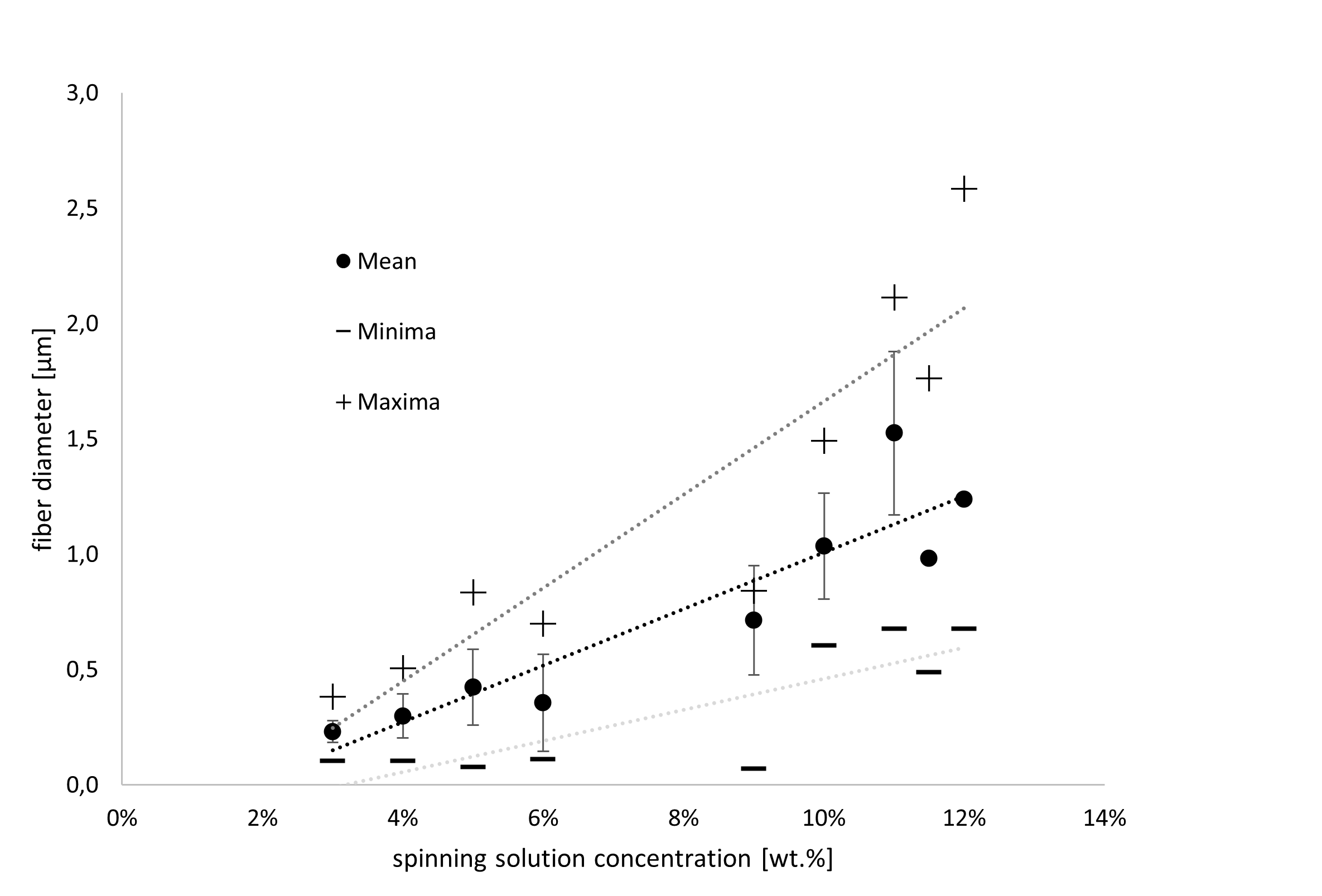
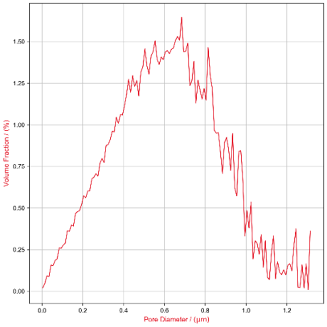
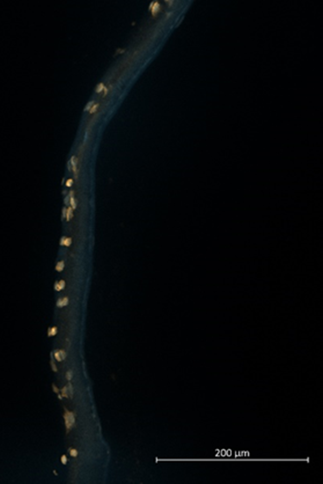
Barrier layer: To create submicroscale pores, the interactions between process and spinning solution parameters as well as fiber diameters were first investigated in detail. The spinning solution concentration (2–15%), the silk fibroin (SF)–polycaprolactone (PCL) ratio (0–100% SF), the influence of salt additives (up to 5 wt-% potassium chloride - KCl), the flow rate (0.5–2 ml/h), and the electric field strength (0–1.5 kV/cm) were systematically varied. It was found that fiber diameters can be specifically reduced, in particular by lowering the flow rate and the spinning solution concentration (Figure 4A) as well as by adding up to 66% SF. Higher SF content and salt additives lead to inhomogeneities in the resulting fiber diameters and shapes, as well as, in some cases, to embrittlement of the membranes. Varying the electric field strength (0–40 kV) also affected the fiber diameters, though only to a very small extent (approx. ± 22 nm); therefore, the preferred solution was selected here based on differences in handling during membrane production. Concentrations below 3% were not spinnable or resulted in irregular strand breaks. Based on these findings, the case group SF:PCL_1:2_3% was selected as the preferred solution. This case group exhibited nearly normally distributed pore sizes with a mean of 613 nm (Figure 4B). Approximately 6% of the pores had a diameter below the target value of 200 nm. To investigate migration behavior, the membranes were mounted in cell culture inserts (Cell-Crown™ inserts) and seeded with gingival epithelial cells for 72 h. After cultivation, cryosections were prepared, and the cytoskeleton and cell nuclei were stained. The barrier function was demonstrated over a period of 72 h (Figure 4C).

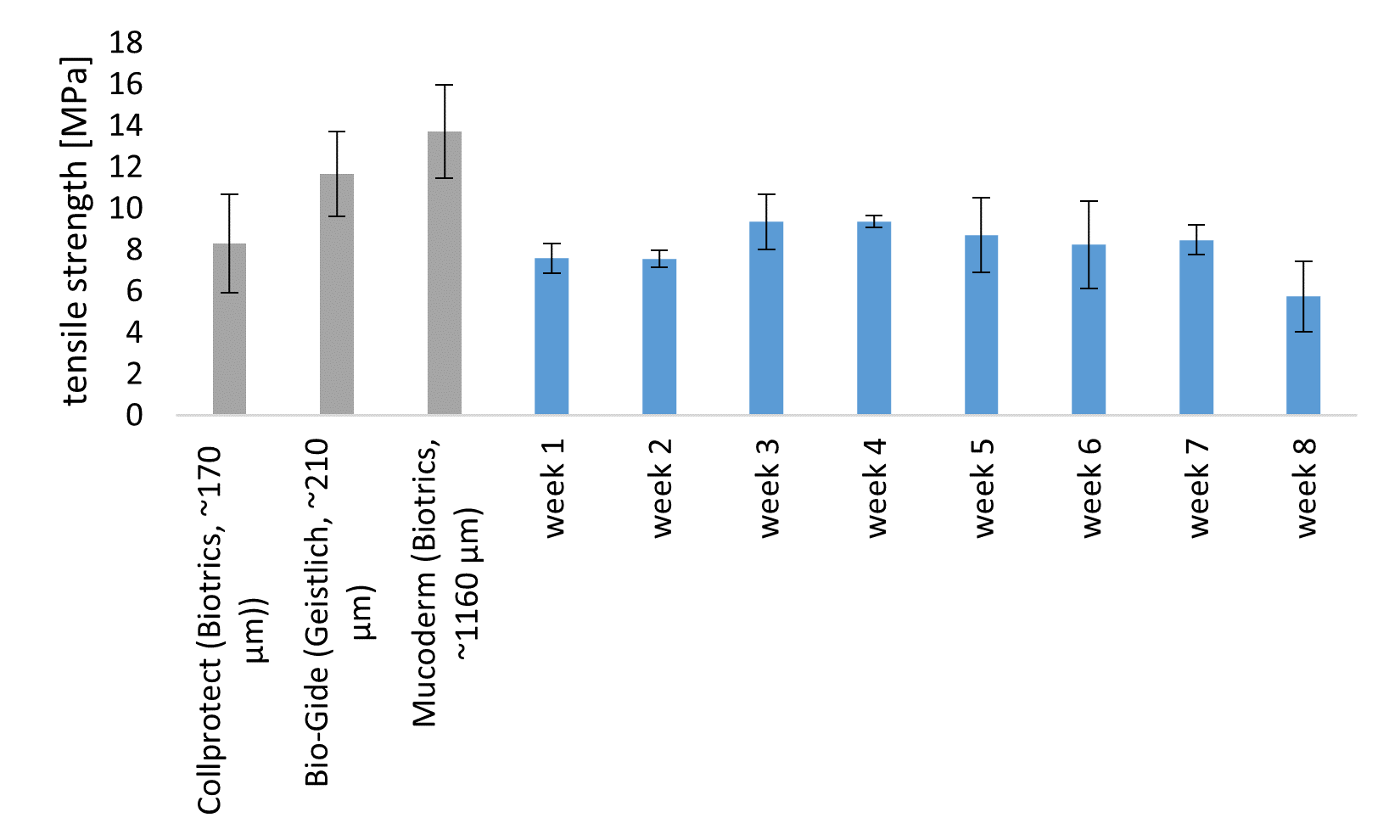
Mineral Layer: To achieve the desired structure of macroporous reinforcement structures, a tricalcium phosphate (TCP)-containing printing paste based on PCL was developed for the FAM. Mineral content of up to 50% was achieved without compromising printability. Using the FAM, constructs with triangular pore geometries were produced, as these offered advantages in terms of surgical handling, particularly with regard to flexibility. The 3D-printed specimens were subjected to enzymatic-catalyzed degradation over an 8-week period and, depending on the degree of degradation, were mechanically characterized in a humid environment and compared with commercially available, non-degraded GBR membranes (BioGuide, CollProtect, Mucoderm). To this end, four test specimens per week were incubated for 8 weeks in a degradation solution (physiological phosphate-buffered saline (PBS) and an enzyme cocktail consisting of protease XIV and lipase). The medium was changed twice each week to prevent the degradation solution from becoming saturated with degradation products. The enzyme concentration was set at 2.5 U/L per enzyme so that both the lipase and protease, considered individually, would reflect the total enzyme concentration in human saliva [9]. This increased enzyme concentration enables rapid degradation while also providing reliable data on the minimum shelf life of the developed GBR membrane in the human oral cavity. Initially, a slight increase in mass and tensile strength in the wet state was observed, which is presumably due to the deposition of salts on the surface (Figure 5). Starting in week 3, a continuous decline in mass and mechanical properties can be observed, although this decline is comparatively minor. Even after 7 weeks, the tensile strength remains in the range of 8 MPa and is thus comparable to the initial strength prior to degradation. Thus, the space-maintaining capacity of the developed GBR membrane was demonstrated for approximately 2 months, which is sufficient for the remodeling of natural bone tissue. The target value of > 5 MPa in a moist environment was maintained throughout the entire degradation period. Furthermore, a comparison was conducted with non-degrading, commercially available GBR membranes, demonstrating the comparability of the developed membrane. In addition, continuous calcium release was detected over the course of the degradation period, which may have a positive effect on bone regeneration.

To evaluate the osteoconductive effect of the mineral layer, the differentiation of bone progenitor cells (mesenchymal stem cells, MSCs) into osteogenic cells was investigated. Based on the staining of calcium deposits in the mineralized tissue (Alizarin Red S staining) and the quantitative analysis of osteogenic marker expression (qPCR of collagen I (Col1), alkaline phosphatase (ALP), osteocalcin (BGLAP), and osteopontin (SPP1)), a positive effect of the membrane developed in the project on bone tissue regeneration was demonstrated. Osteogenic induction was achieved by adding dexamethasone, β-glycerophosphate, and ascorbic acid for 28 days. Differentiation was controlled using MSCs without any material influence.

Summary: As a result of the OsteoMe project, three membrane layers were developed, each of which fulfills essential aspects of the requirements profile for GBR membranes in dentistry. Depending on the acidity of the chitosan as well as the contact time and pressure, the mucoadhesive layer develops adhesive forces in the range of 1 to 4 N, which are comparable to those of established fibrin glues. This enables the membrane to be applied to the defect site in a position-stable manner without the need for additional sutures or pins. The barrier layer is designed with open pores, allowing for the exchange of nutrients and signaling molecules, which can positively influence regeneration. At the same time, it was shown that the pore size is small enough to act as a barrier to rapidly proliferating oral mucosal cells, thereby providing the slow-growing bone tissue cells with sufficient time for regeneration. The 3D-printed mineral layer exhibits sufficiently high mechanical properties in simulated saliva to ensure a space-maintaining function. The tensile strengths achieved are comparable to those of commercially available GBR membranes and show virtually no decline during enzymatic-catalyzed degradation over eight weeks. Furthermore, it was demonstrated that the mineral layer exhibits osteoinductive properties and can thus actively support bone regeneration. Finally, methods were developed to ensure a delamination-free bonding of the individual layers. This allows the layers to be flexibly combined with one another, thereby expanding the range of possible applications.


Acknowledgments: The IGF project 01IF22810N of the Forschungskuratorium Textil e.V. research association was funded by the Federal Ministry for Economic Affairs and Energy through the DLR Project Management Agency as part of the Program for the Promotion of Industrial Collaborative Research (IGF), pursuant to a resolution of the German Bundestag.


Quellenverzeichnis:

[1]         CHOLMAKOW-BODECHTEL, Constanze: Fünfte Deutsche Mundgesundheitsstudie (DMS V). JORDAN, Andreas Rainer (Hrsg.); MICHEELIS, Wolfgang (Hrsg.). Köln : Deutscher Zahnärzte Verlag DÄV, 2016

[2]         CHA, Hyun-Suk ; KIM, Ji-Wan ; HWANG, Jong-Hyun ; AHN, Kang-Min: Frequency of bone graft in implant surgery. In: Maxillofacial plastic and reconstructive surgery 38 (2016), Nr. 1, S. 19

[3]         MOY, Peter K. ; AGHALOO, Tara: Risk factors in bone augmentation procedures. In: Periodon-tology 2000 81 (2019), Nr. 1, S. 76–90

[4]         KNÖFLER, Wolfram ; BARTH, Thomas ; GRAUL, Reinhard ; KRAMPE, Dietmar: Retrospective analysis of 10,000 implants from insertion up to 20 years-analysis of implantations using aug-mentative procedures. In: International journal of implant dentistry 2 (2016), Nr. 1, S. 25

[5]         CABALLÉ-SERRANO, Jordi ; MUNAR-FRAU, Antonio ; ORTIZ-PUIGPELAT, Octavi ; SOTO-PENALOZA, David ; PEÑARROCHA, Miguel ; HERNÁNDEZ-ALFARO, Federico: On the search of the ideal barrier membrane for guided bone regeneration. In: Journal of clinical and experi-mental dentistry 10 (2018), Nr. 5, e477-e483

[6]         JIMÉNEZ GARCIA, J. ; BERGHEZAN, S. ; CARAMÊS, J. M. M. ; DARD, M. M. ; MARQUES, D. N. S.: Effect of cross-linked vs non-cross-linked collagen membranes on bone: A systematic re-view. In: Journal of periodontal research 52 (2017), Nr. 6, S. 955–964

[7]         BUTENSCHÖN, Sina: Prävalenz periimplantärer Entzündungen bei teilbezahnten Patienten nach einer minimalen Beobachtungsdauer von 10 Jahren - eine retrospektive Querschnittsstu-die. Göttingen, Georg-August-Universität zu Göttingen. Dissertation. 2019

[8]         RAKIC, Mia ; GALINDO-MORENO, Pablo ; MONJE, Alberto ; RADOVANOVIC, Sandro ; WANG, Hom-Lay ; COCHRAN, David ; SCULEAN, Anton ; CANULLO, Luigi: How frequent does peri-implantitis occur? A systematic review and meta-analysis. In: Clinical oral investigations 22 (2018), Nr. 4, S. 1805–1816

[9]         CHAUNCEY, Howard Haskell. The chemical composition of human sal

AutorInnen: Lukas Benecke Claudia Dietze Ina Prade Michael Meyer Chokri Cherif

Technische Universität Dresden
Fakultät Maschinenwesen
Institut für Textilmaschinen und Textile Hochleistungswerkstofftechnik (ITM)
01062 Dresden

https://tu-dresden.de/mw/itm


FILK Freiberg Institute gGmbH
Meißner Ring 1-5
09599 Freiberg

Deutschland

https://www.filkfreiberg.de/

More entries from TU Dresden, Institut für Textilmaschinen und Textile Hochleistungswerkstofftechnik ITM

05.02.2025

Integral flachgestrickte Drucksensoren für smart Textiles

Gestricke & Gewirke Sensorik Smart Textiles Tests

Zusammenfassung

Im IGF-Projekt 21990 BR1 wurde das „Textiles Smart-Skin-3D-System (S3D)“ entwickelt – ein innovatives, flachgestricktes Sensorsystem, das Druck- und Näherungsmessungen nahtlos in textile Produkte integriert. Ziel war es, flexible und robuste Sensorik bereits im Herstellungsprozess einzubetten und so die Komplexität sowie potenzielle Schwachstellen herkömmlicher Mehrkomponentensysteme zu vermeiden. Hierzu wurden komplexe 3D-gestrickte Strukturen realisiert, die leitfähige Sensorgarne und gezielt eingearbeitete dielektrische Materialien wie silikonbasierte Inserts nutzen, um kapazitive Messprinzipien anzuwenden.

Die Optimierung von Garnauswahl und Strickparametern ermöglichte eine präzise Erfassung von Druckkräften und Annäherungen. Als Demonstrator wurde ein vollständig integrierter Sensorhandschuh mit 13 Sensorflächen entwickelt, der Greif- und Haltekräfte misst. Zyklische elektromechanische Prüfungen bestätigten ein stabiles Sensorverhalten. Insbesondere zeigte die Variante mit einem 1 mm starken Dielektrikum optimale Übertragungscharakteristika, geringe Hysterese und eine Sensordrift im akzeptablen Rahmen. Zusätzlich erbrachte ein textilbasierter Näherungssensor zuverlässige Messwerte für Abstände bis zu 120 mm.

Die Ergebnisse belegen das Potenzial flachgestrickter Sensoren als integraler Bestandteil smarter, tragbarer Textilien – mit Anwendungsmöglichkeiten in Telerehabilitation, Medizintechnik, Arbeitsschutz und weiteren Digitalisierungsbereichen.

Summary

In the IGF project 21990 BR1, the “Textiles Smart-Skin-3D-System (S3D)” was developed – an innovative, flat-knit sensor system that seamlessly integrates pressure and proximity measurements into textile products. The aim was to embed flexible and robust sensor technology into the manufacturing process, thereby avoiding the complexity and potential weaknesses of conventional multi-component systems. To achieve this, complex 3D-knit structures were created using conductive sensor yarns and strategically incorporated dielectric materials, such as silicone-based inserts, to implement a capacitive sensing approach.

Optimizing yarn selection and knitting parameters enabled the precise detection of pressure forces and proximity. A demonstrator in the form of a fully integrated sensor glove with 13 sensing areas was developed, capable of measuring gripping and holding forces. Cyclic electromechanical tests confirmed stable sensor performance. In particular, the variant with a 1 mm thick dielectric exhibited optimal transfer characteristics, low hysteresis, and acceptable sensor drift. Additionally, the textile-based proximity sensor reliably measured distances of up to 120 mm.

The results demonstrate the potential of flat-knit sensors as an integral component of smart, wearable textiles with applications in telerehabilitation, medical technology, occupational safety, and other digitalization sectors.

Bericht

Einleitung

Vor dem Hintergrund globaler Megatrends wie der Digitalisierung in der Medizin bestehen für die Textilindustrie große Chancen, vom erwarteten weiteren Wachstum von am Körper tragbaren, flexibel einsetzbaren und computergestützten Systemen zu profitieren. Zu dieser neuen Geräteklasse, den sogenannten Wearables, gehören Textilien, die über die klassischen Funktionen von Bekleidung oder beispielsweise Bandagen hinaus mit elektronischen Zusatzfunktionen ausgestattet sind. Da Textilien häufig die Schnittstelle zwischen dem Menschen und seiner Umwelt darstellen, sind sie prädestiniert, auch bei der Digitalisierung menschlicher Wahrnehmungen und Fähigkeiten (z. B. Bewegungen, Haptik etc.) und umgekehrt der Rückkopplung von der virtuellen in die analoge Welt eine entscheidende Brückenfunktion zu übernehmen und so als künstliche Haut (bzw. Smart Skin) bestehende optische und akustische Schnittstellen zu ergänzen.

Ein Bereich in dem smarte Textilien einen großen Zugewinn nützlicher Informationen bereitstellen, ist die Medizin und Rehabilitationstechnik. Vor dem Hintergrund einer alternden Bevölkerung und damit einhergehend einer hohen Belastung medizinischer Versorger, die unter gleichzeitigem Personalmangel leiden, ist nicht immer ein ausreichendes Angebot in erreichbarer Nähe realisierbar. Vor allem im Bereich der medizinischen Folgebehandlungen für Physiotherapie einhergehend mit langen Transportwegen oder fehlender Transportfähigkeit des Patienten kann dies zu Heilungsverlangsam oder sogar -verhinderung führen. Eine Unterstützung von Patienten durch einen medizinischen Laien (Familienangehörige, Bekannte etc.) mit einem geringfügigen Lernaufwand soll durch den in diesem Projekt entwickelten Handschuh ermöglicht werden. Dieser ermöglicht die Überwachung von Greif- und Haltebewegungen sowie Feedback zur Korrektur. In der Telerehabilitation gibt es keine vergleichbaren Systeme, die autonom ohne Experteneinsatz arbeiten [1, 2]. Das Projekt fokussierte auf die Entwicklung multifunktionaler Druck-/ Näherungssensorik durch flachstricktechnische Verfahren. Diese ermöglichen die kostengünstige Integration in Funktionsbekleidung, aber auch in Roboterkomponenten.

Zielsetzung und Lösungsweg

Das Ziel des IGF-Forschungsprojekts war die Entwicklung, Charakterisierung und Erprobung textilbasierter Drucksensoren, die mittels Flachstricktechnik in einen Handschuh integriert werden sollten um die aufgebrachte Kraft auf den Fingergliedern und dem Handballen zu überwachen. Es wurden flächenbasierte, gestrickte Sensorkonzepte mit einem kapazitiven Messprinzip verfolgt. Die entwickelten Sensoren wurden mittels zyklischer elektromechanischer Druckprüfungen untersucht und eine Vorzugsvariante der Sensoren zur Integration in einem Funktionsdemonstrator ermittelt. Weiterhin wurden kapazitive Näherungssensoren entwickelt und evaluiert.

Ergebnisse

Entwicklung der gestrickten Drucksensoren

Für die Entwicklung der Sensoren wurde die Umsetzung eines kapazitiven Drucksensors mithilfe von Flachstricktechnik verfolgt. Die Vorteile kapazitiver Sensoren gegenüber resistiver Sensoren liegen in ihrer Unempfindlichkeit gegenüber Temperatur [3], was in einer körpernahen Anwendung von Vorteil ist. Der einfachste Aufbau eines Kondensators ist der Plattenkondensator. In diesem Aufbau sind zwei parallele Platten durch ein Dielektrikum getrennt. Durch das Aufbringen einer Druckkraft F auf diese Platten und damit ein Zusammendrücken des Dielektrikums mit der Dielektrizitätskonstante  ε ändert sich der Plattenabstand d und somit die Kapazität C wie in Abbildung 1 gezeigt. Hier wird deutlich, dass die Kapazitätsänderung ∆C indirekt proportional zur Änderung des Plattenabstands ∆d, die wiederum abhängig ist von der induzierten Kraft, dem E-Modul E und den geometrischen Maßen des Plattenkondensators mit b = Breite und l = Länge.

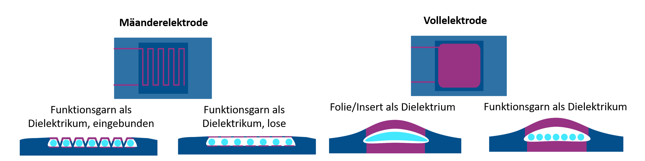
Für den Aufbau der gestrickten kapazitiven Sensoren wurden verschiedene Konzepte erstellt, die in Abbildung 2 dargestellt sind. Anhand einer systematischen Variantenbewertung nach ergonomischen, stricktechnischen, sensortechnischen Anforderungen und praktischer Versuchstests wurde eine Sensorvariante mit einem Insert als Dielektrikum und einer vollflächigen Elektrode aus leitfähigem Garn als Vorzugsvariante gewählt und zu einer Handschuhfinger gleichenden Doppelschlauchstruktur erweitert.

Zur Auswahl des Elektrodengarns wurden Vorversuche durchgeführt um die stricktechnische Eignung der teilweise anspruchsvoll zu verarbeitenden Garne auf Stahl- und Silberbasis zu bewerten. Hierbei wurden Garne von Statex (Shieldex® 235 f 36dtex Z130), Amann (Steel-tech® 100 tex 93, Silver-tech+® 150 tex 22) und Bekaert (Bekinox® VN 14.1.9.100Z) genutzt. In diesen Vorversuchen erwies sich Silver-tech+® 150 als Vorzugsvariante, da es sehr gut mit dem umgebenden Basismaterial aus Umwindegarn (Tencel CV Nm40 mit PA6.6 78/78f23/1) fertigungstechnisch kompatibel war.

Herstellung der Sensoren

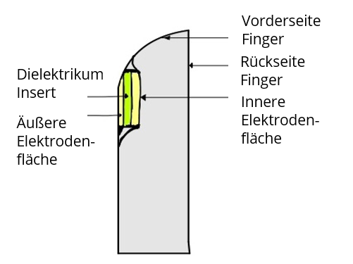
Ziel des Projekts war die Herstellung eines Sensorhandschuhs mittels Flachstricktechnik, eine Strickmethode, die die Möglichkeit bietet Fully Fashioned Artikel in einem Arbeitsschritt herzustellen, wodurch komplizierte gestrickte Flächen endkonturnah hergestellt werden können. Um ein höchstmöglich automatisiert herstellbares Produkt zu entwickeln wurde der Drucksensor mit einem Fokus auf Vermeidung nachfolgender Konfektionierungsschritte entwickelt. Daher wurde der Drucksensor als eine Doppelschlauchstruktur konzeptioniert. Diese wird durch zwei Elemente geformt: Zum einen durch die Tasche des Sensors, zum anderen durch einen Fingerling, der eine Tragbarkeit des Sensors ermöglicht. In Abbildung 3 ist der Aufbau schematisch dargestellt. Im Sensorbereich ergibt sich daher ein dreilagiges Doppelschlauch-gestrick. Das umfasst die äußere sowie innere Elektrode und die Rückseite des Fingers. Das Dielektrikum wird durch ein Insert, welches während des Strickprozesses eingelegt wird, gebildet. Diese Variante des Konzeptes ermöglicht eine weitestgehend automatisierte Fertigung des Handschuhs an der Flachstrickmaschine ohne nachgelagerte Konfektionsschritte. Für die Einbringung des Dielektrikums ist eine Unterbrechung des Strickprozesses erforderlich.

Validierung der Sensoren

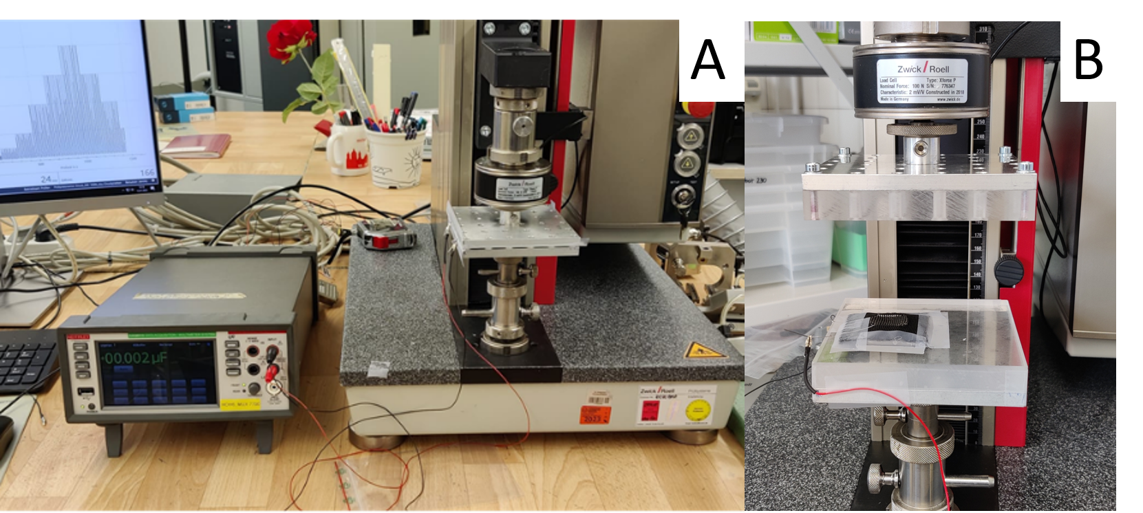
Die gestrickten kapazitiven Sensoren wurden auf ihre Eignung als Drucksensor in zyklischen elektromechanischen Messungen überprüft. Der Versuchsaufbau mit Mess- und Versuchsgeräten sowie der Prüfablauf sind in Abbildung 4 dokumentiert. Um den Einfluss des Dielektrikums zu untersuchen, wurden Sensoren mit einem 2 mm und einem 1 mm starken silikonbasierten Dielektrikum hergestellt. Aus den ermittelten Daten wurden das Übertragungsverhalten (als Zusammenhang zwischen Kompressionskraft und Sensorsignal), die Sensordrift (als Signalwerte bei Entlastung der Sensoren) und die Hysterese (als maximale Differenz zwischen Be- und Entlastungskurve über den Messbereich) berechnet (siehe Abbildung 5).

Es zeigte sich, dass beide Varianten ein stabiles Sensorverhalten aufweisen, wobei die Sensorvariante mit einem 1 mm starken Dielektrikum bessere Ergebnisse im Übertragungsverhalten und in Hysterese zeigte. Die Sensordrift lag hier etwas höher, lag aber bei beiden Varianten unter 5 % und damit in einem, für praktische Anwendungen dieser Technologie, akzeptablen Bereich. Dieser Versuch zeigte, dass das Dielektrikum einen entscheidenden Einfluss auf das Sensorverhalten hat und dieses durch die relativ kleine Anpassung des Insertmaterials für verschiedene Messbereiche und -sensitivitäten angepasst werden kann. Weitere Ausführungen, Ergebnisse und Diskussionen können aus der Publikation in [4] entnommen werden.

Näherungssensor

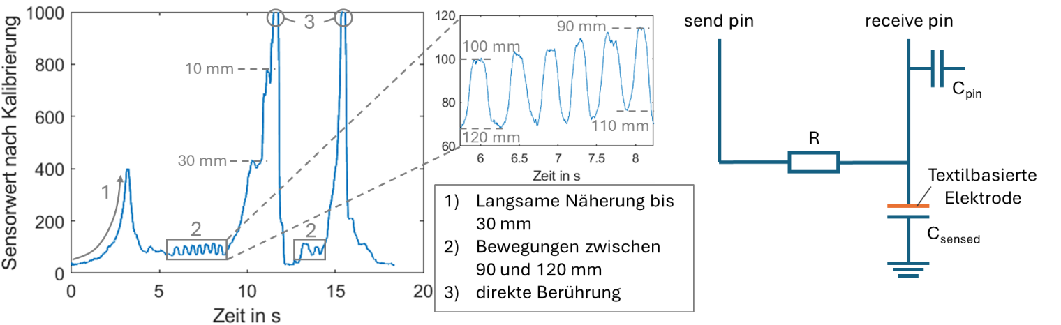
Das Konzept für die textile Näherungssensorik wurde mit einer einzelnen textilen gestrickten Elektrode und einem Arduino Uno umgesetzt. Für die Versuchsdurchführung wurde eine menschliche Hand als zu erfassendes Objekt an den Sensor geführt und der Abstand zwischen Hand und Sensor gemessen. In Abbildung 6 sind das Sensorsignal und korrelierte Abstände der Hand dazu gezeigt, sowie das Schaltbild dargestellt. Hierbei konnten Abstände von bis zu 120 mm zur Hand noch erfasst werden mit einer guten Signalstabilität, sodass hier eine Quantifizierung des Abstands denkbar ist.

Demonstrator

Die Vorzugsvariante für den Druck- und Näherungssensor wurde übertragen auf einen vollständig gestrickten und integral gefertigten Handschuh mit 13 Sensoren, wobei 2 Sensorflächen für Daumen, 3 Sensorflächen für Zeige- und Mittelfinger und 5 Sensorflächen auf der Handfläche für die Erfassung von Kräften realisiert wurden. Der finale Funktionsdemonstrator ist in Abbildung 7 gezeigt. Die elektrischen Zuleitungen wurden für diesen FD manuell realisiert. Eine sensorische Funktionalisierung des Ringfingers und des kleinen Fingers war durch die begrenzte Anzahl an Fadenführern innerhalb der Strickmaschine nicht möglich (max. 13 Sensorflächen). Die Signale der einzelnen Sensoren wurden mittels eines RaspberryPi 5 und einer dafür entwickelten Software ausgewertet. In verschiedenen Greiftests wurden die Sensoren validiert. Bei allen funktionsfähigen Sensoren konnte ein verlässlicher Anstieg des Signals bei Kompression erfasst werden.

Zusammenfassung und Ausblick

Die Verwendung textiltechnischer Lösungen zur Überwachung des menschlichen Körpers und der auf ihn wirkenden Lasten ist ein vielversprechendes Forschungsfeld, das Anwendungen in der Physiotherapie, im Arbeitsschutz und in der Digitalisierung von Arbeitsprozessen ermöglicht. Im Rahmen dieses Projekts lag der Fokus auf der Entwicklung und Integration von Druck- und Näherungssensoren in textile Strukturen. Dabei wurden innovative textilbasierte Ansätze verfolgt, insbesondere die Herstellung vollständig textilintegrierter Sensoren im Fully-Fashioned-Verfahren. Im Gegensatz zu herkömmlichen Systemen, die oft aus vielen Einzelkomponenten bestehen und dadurch Schwachstellen aufweisen, bieten textilbasierte Sensorsysteme eine höhere Kompatibilität mit textilen Basissystemen und eine höhere Flexibilität. Die in dieser Arbeit entwickelten Sensoren sind vielseitig einsetzbar und können in zahlreiche textile Strukturen, und vor allem gestrickter Strukturen, diverser Form und Größe übertragen werden.

Unter Beachtung industrienaher Anforderungen, die zusammen mit den am Projekt beteiligten Industriepartnern festgelegt wurden, wurden verschiedene Konzepte für Druck- und Näherungssensoren für einen Sensorhandschuh unter Nutzung von Flachstricktechnik entwickelt. Die bevorzugte Lösung für gestrickte Druck- und Näherungssensoren basiert auf einem Doppelschlauchgestrick, das einen flexiblen Plattenkondensator darstellt. Diese Sensoren bestehen aus Elektroden aus leitfähigem Garn und einem weichen Material, beispielsweise Silikon, das als Dielektrikum dient. Dadurch, dass das Material für das Dielektrikum flexibel gewählt werden kann, sind Messbereich und -verhalten auch für andere Anwendungen mit diesem Konzept einfach zu variieren. Für die Druckmessung wurde das Ansprechverhalten der entwickelten Sensoren eingehend getestet, und ihre Stabilität analysiert und ein funktionsgerechtes Messverhalten der Sensoren im Messbereich 0 bis 10 N festgestellt.

Die Vorzugsvariante der Sensoren wurde in einem Funktionsdemonstrator mit 13 Sensorflächen umgesetzt. Dies sollte in weiteren Arbeiten um 6 weitere Sensorflächen für die einzelnen Fingergelenke von Ring- und kleinem Finger ergänzt werden. Die Anzahl der Sensorflächen war in diesem Projekt durch die Anzahl der verfügbaren Fadenführer begrenzt. Weiterhin sollte das Einlegen des dielektrischen Inserts stärker automatisiert werden um die Zeit, die benötigt wird um die Drucksensorhandschuhe zu stricken, reduziert wird.

Danksagung

Das IGF-Vorhaben 21990 BR der Forschungsvereinigung Forschungskuratorium Textil e.V., Reinhardtstr. 12-14, 10117 Berlin wurde über die AiF im Rahmen des Programms zur Förderung der industriellen Gemeinschaftsforschung und -entwicklung (IGF) vom Bundesministerium für Wirtschaft und Klimaschutz aufgrund eines Beschlusses des Deutschen Bundestages gefördert.

Die Autoren danken den genannten Institutionen für die Bereitstellung der finanziellen Mittel. Der Forschungsbericht und weiterführende Informationen sind am Institut für Textilmaschinen und Textile Hochleistungswerkstofftechnik der TU Dresden erhältlich.


Literatur


[1]   K. Ettle et al., "Telepräsenzroboter für die Pflege und Unterstützung von Schlaganfallpatientinnen und -patienten (TePUS) im Regierungsbezirk Oberpfalz: DeinHaus 4.0," Regensburg, Jun. 2020. Accessed: Nov. 30 2020.

[2]   K. Berkenkamp, "Telerehabilitation in der Schlaganfallversorgung – Einflussfaktoren auf Adoption und Akzeptanz von klinisch tätigen Ärzten und Therapeuten," 2020.

[3]   J. Mersch, C. A. G. Cuaran, A. Vasilev, A. Nocke, C. Cherif, and G. Gerlach, "Stretchable and Compliant Textile Strain Sensors," IEEE Sensors J., vol. 21, no. 22, pp. 25632–25640, 2021, doi: 10.1109/JSEN.2021.3115973.

[4]   S. Fischer, C. Böhmer, S. Nasrin, C. Sachse, C. Cherif. Flat-Knitted Double-Tube Structure Capacitive Pressure Sensors Integrated into Fingertips of Fully Fashioned Glove Intended for Therapeutic Use. Sensors 2024, 24, 7500. https://doi.org/10.3390/s24237500


AutorInnen: Carola Bömer

Technische Universität Dresden
Fakultät Maschinenwesen
Institut für Textilmaschinen und Textile Hochleistungswerkstofftechnik (ITM)
01062 Dresden

https://tu-dresden.de/mw/itm

More entries from TU Dresden, Institut für Textilmaschinen und Textile Hochleistungswerkstofftechnik ITM

18.10.2022

Development of Textile Structures with Material-Intrinsic Shape Changing Capabilities for Regenerative Medicine (TexMedActor)

Garne Gewebe Nachhaltigkeit Technische Textilien Medizin

Zusammenfassung

In the IGF project 21022 BR/1 "TexMedActor", fabrics based on shape memory or electroactive yarns were developed which are capable of enclosing defects in hollow organs on the one hand and stimulating cells by micro-movements on the other. For this purpose, influences of spinning process and material composition on the shape memory behavior of TPU-based yarns were characterized and, in particular, the activation temperature was adjusted to values of the body core and body surface temperature. Furthermore, piezoelectric PVDF yarns were developed whose proportion of polar crystal phases was significantly increased by the spinning parameters and post-treatment, which also increased the piezoelectric behavior of the material. This allowed dynamic changes in pore size to be demonstrated in situ, which can have a stimulating effect on cells. With a new process and a new product group (textiles with intrinsic, active shape-changing capability), the results offer high innovation potential not only for medical devices, but also for a wide range of lucrative applications in a variety of niches, such as sports textiles and filter textiles. Furthermore, these can be used as a basis for the development of extracorporeal medical products such as compression textiles, bandages and orthoses.

Bericht

Introduction and Objective

In Germany, both demographic changes in society and injuries resulting from trauma are leading to a high proportion of people with cardiovascular diseases or injuries to vessels and internal organs requiring treatment. Treatment of injuries to internal organs, vessels, or nerves usually requires complex procedures (anastomoses) that involve elaborate fixation and suturing. These complicated and elaborate procedures are often associated with long procedure times, which in turn directly correlate with increased complication rates [1-3]. Tubular plastic implants are increasingly being developed to bridge such defects. These single material structures do not allow tissue/ cell ingrowth. Therefore, they run counter to the concept of regenerative medicine, which aims to restore body tissues and cells. In addition, when the defects are filled, regeneration is often disturbed due to the structural-mechanical properties that are not adapted to biomechanics. Furthermore, the lack of interconnectivity of the pore spaces of the replacement structures prevents the cell ingrowth, cell growth, nutrient supply and the removal of metabolic products.

In the context of in vitro tissue engineering, in addition to static cell culture systems, dynamic systems are also being developed. These are based, for example, on continuous or pulsating fluid flows or on a cyclic stretching of a clamped cell support system or substrate [4]. However, a replication of natural mechanical growth stimuli is not possible with such bioreactor systems because, especially in larger structures, there is a locally increased flow velocity along the largest pores or only an overflow of the entire cell support system. Additionally, undesirable stress peaks and undefined distortions occur in the region of the clamps and supports in mechanically stimulated systems.

Since the native structure of the four most important tissue types (connective and supporting tissue, nervous, muscular and epithelial tissue) from which organs, such as bones, blood vessels, muscles, tendons and ligaments, are formed, consists of fiber-like constructs, these can be particularly well biomimicked with textile structures. With the help of pre-designed fiber arrangements, three-dimensional, complex geometries with interconnecting pore spaces can be built up. The cells can use these structures to orient themselves in their growth direction [5]. Therefore, fiber-based high-tech structures are particularly predestined to overcome the limitations of currently available implants.

Therefore, within the framework of the IGF research project TexMedActor (21022 BR/1) novel textile structures with material-intrinsic shape changing capabilities were developed for regenerative medicine with a variety of different application fields, especially anastomosis. The concept pursued envisages the textile-technological realization of structures with a shape memory effect. The textiles should be able to assume predetermined geometries in order to adapt interactively to defects and to simplify complex interventions to bridge or support defects in internal organs like vessel and nerves. Furthermore, these textiles are intended to enable electromechanical stimulation for the actively targeted stimulating of cell growth. In this way, regeneration is accelerated or even made possible in the first place, since the necessary stimuli for tissue- and cell-adapted growth stimulation are lacking, especially in the case of body tissues with weak or no blood supply, such as cartilage, tendons, ligaments, or in the case of wound healing disorders or chronic wounds. Furthermore, novel bioreactors based on the intrinsic properties of the textile structures will be developed, which use the mechanism of action for electromechanical stimulation to uniformly stimulate the cells at each site even in highly complex and large-scale cell carrier structures. Here, the mechanical stimuli originate from the material itself. This material-intrinsic stimulation represent a new method for optimal cell cultivation, by stimulating cell on the textile cell carrier structures without externally applied fluid flows or mechanical deformation. This is intended to overcome two recognized medical technology problems: 1) complicated, costly operations on internal organs, vessels or nerves that are difficult or impossible to perform with minimally invasive procedures, and 2) lack of tissue- and cell-adapted stimuli for promotion of growth in previously used replacement structures and materials as well as currently available dynamic cell culture systems.

Acknowledgement

The IGF project 21022 BR/1 of the Research Association Forschungskuratorium Textil e.V. was funded by the Federal Ministry of Economics and Climate Protection via the AiF within the framework of the program for the promotion of joint industrial research (IGF) on the basis of a resolution of the German Bundestag. We would like to thank the above-mentioned institutions for providing the financial resources. Furthermore, we want to thank the member of the “Projektbegleitender Ausschuss” (project accompanying committee) for their support during the project.

AutorInnen: Benecke, Lukas; Aibibu, Dilbar; Cherif, Chokri

Technische Universität Dresden
Fakultät Maschinenwesen
Institut für Textilmaschinen und Textile Hochleistungswerkstofftechnik (ITM)
01062 Dresden

More entries from TU Dresden, Institut für Textilmaschinen und Textile Hochleistungswerkstofftechnik ITM