
OsteoMe: Multilayered GBR Membranes with Mucoadhesive and Osteoinductive Properties for Bone Augmentation
Abstract
The OsteoMe project, funded as part of the Joint Industrial Research (IGF) program, focuses on the development of a multilayer membrane that combines mucoadhesive, cell-excluding, and space-maintaining properties through a material- and structurally graded layered structure. With this property profile, combined with the use of bioresorbable materials, the project aims to meet all requirements for guided bone regeneration (GBR) membranes in dentistry and to specifically improve upon or surpass existing solutions. For manufacturing, electrospinning was combined with 3D printing and surface functionalization. As a result, three membrane layers were developed that individually represent specific aspects of the property profile and can be flexibly combined with one another.
Report
Introduction: The causes of tooth loss are very diverse, including trauma, tumor removal, tooth decay, and—very commonly—periodontitis (bacterial inflammation of the gums and jawbone). In Germany, approximately 50–60% of the population suffers from moderate to severe periodontitis, with the incidence increasing with age; consequently, a growing demand for artificial teeth is expected in the future [1]. In addition to the root cavity resulting from tooth loss, the causes mentioned above can also lead to damage to larger areas of the jawbone or necessitate their removal. Furthermore, bone resorption occurs due to the lack of mechanical stress on the jaw, making it impossible to place an implant without prior reconstruction of the bone tissue. Since an implant must be firmly anchored in the jawbone, prior bone augmentation is therefore absolutely necessary in these cases to provide patients with optimal care. In Germany, over 50% of dental implants placed require such augmentation [2–4]. In this treatment approach, a bone substitute material is filled into the cavity and covered with a GBR membrane. The membrane is essential for the success of the treatment, as its primary function is to prevent rapidly proliferating soft tissue cells from invading the cavity. Currently, both non-resorbable (e.g., PTFE, titanium) and resorbable (e.g., collagen) GBR membranes are used; however, they can only partially meet the required performance criteria. Resorbable membranes tend to be preferred because they do not need to be removed in a second surgical procedure—which could jeopardize treatment success—and carry a lower risk of infection. However, due to their lack of primary stability, their mechanical properties do not yet allow them to maintain space in a moist environment. For this reason, non-resorbable GBR membranes continue to be used as well [5, 6]. A GBR membrane that combines resorbability with space-maintaining properties in a way that meets clinical requirements does not yet exist, a fact underscored by high revision rates of approximately 20% [7, 8].
The IGF OsteoMe project aimed to develop novel, multilayer GBR membranes for bone augmentation with a complex, application-oriented property profile. To this end, the project partners ITM and FILK combined their complementary expertise in the development of electrospun and additive-manufactured (Fiber Additive Manufacturing, FAM) biopolymer structures, the functionalization of chitosans, and cell biological characterization. The property profile, which had been developed in advance through discussions with clinicians and industry representatives, was to be realized through a three-layer membrane structure consisting of a mucoadhesive layer for positional stabilization, a cell-excluding barrier layer, and a structuring, mineralized layer with defined porosity and mineral gradients to biomimetically replicate the bone-tissue interface and provide space-maintaining properties. Based on the materials polycaprolactone (PCL), silk fibroin (SF), and chitosan (Ch), simulation-based fabrication strategies were investigated that enable the targeted adjustment of mechanical, functional, and biological properties.
Results:
Mucoadhesive layer: Mucoadhesive materials allow for prolonged retention on mucous membranes and are therefore of interest for numerous medical applications. Chitosan is considered a promising mucoadhesive biopolymer due to its positive charge and the resulting interactions with negatively charged mucins. To further improve adhesion, the covalent binding of thiol-containing compounds was investigated, as these additionally enable the formation of disulfide bonds with mucin.
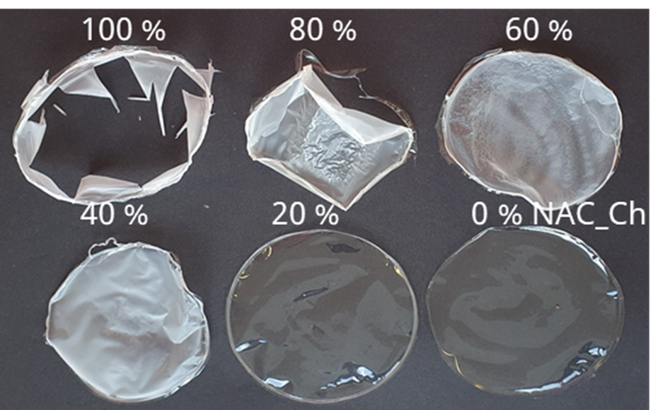
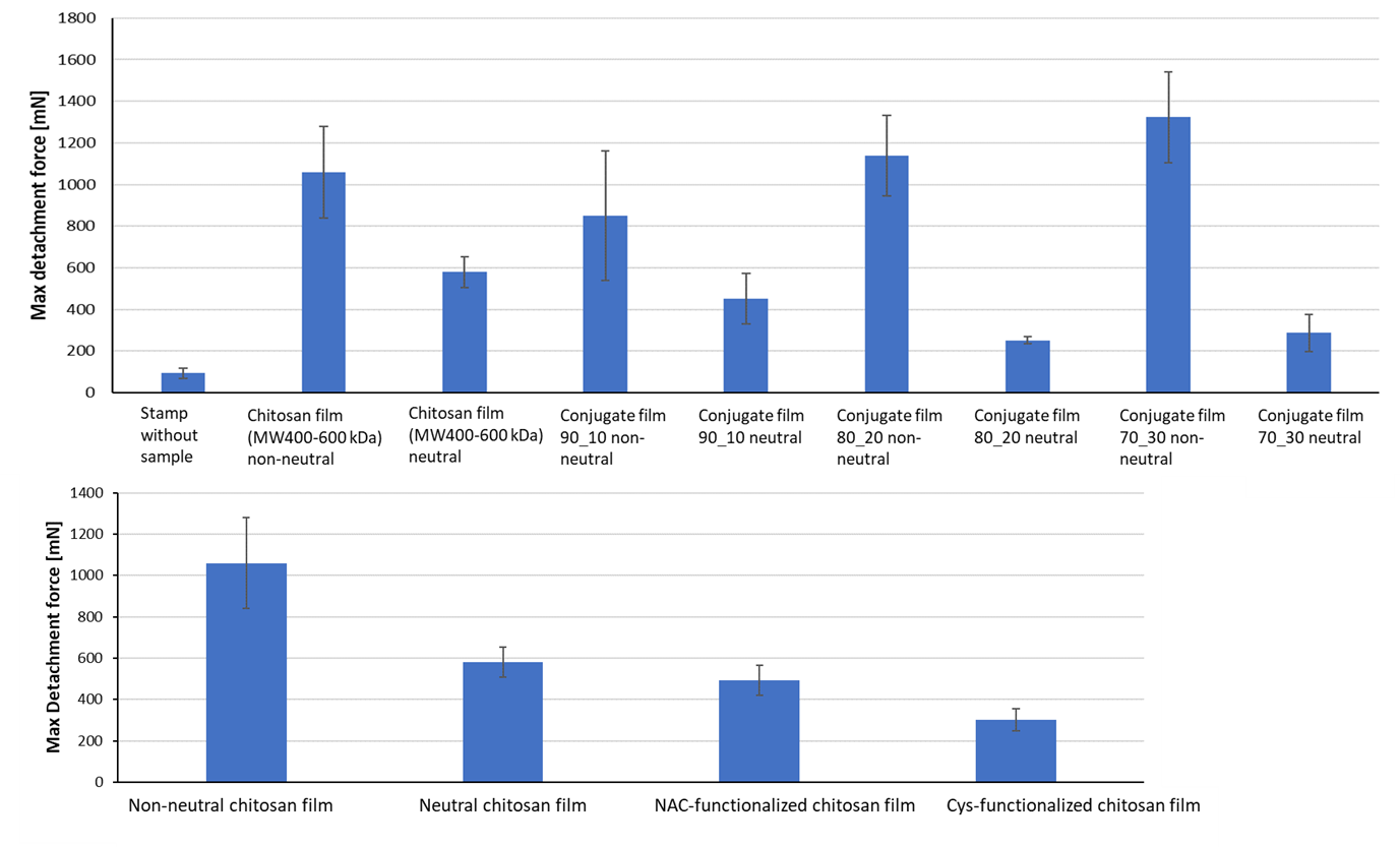
Functionalization was carried out via carbodiimide (EDC/NHS)-mediated coupling of N-acetylcysteine (NAC) or cysteine to chitosan with a degree of deacetylation exceeding 90%. Both the direct modification of chitosan films and the derivatization of dissolved chitosan were investigated. While the functionalization of films was complicated by the limited accessibility of the amino groups within the solid polymer matrix and resulted in an inhomogeneous distribution of thiol groups, the modification of dissolved chitosan enabled the preparation of reproducible conjugates with high thiol contents. The optimized chitosan conjugate achieved a total thiol content of approximately 878 µmol/g, with about 311 µmol/g present as free thiol groups. Due to the high reactivity of the thiol groups, a loss of approximately 20% was observed within two weeks during storage in air; therefore, storage under oxygen-free conditions is recommended. To produce mechanically stable membranes, the addition of at least 70 wt% unmodified chitosan was required (see Figure 1).
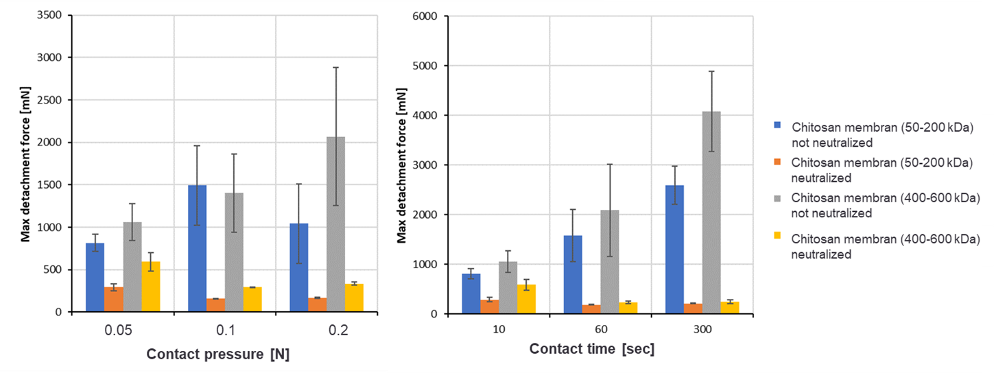
The mucoadhesive properties were investigated using a specially developed test setup on a texture analyzer, with porcine intestinal mucosa serving as the model substrate. The adhesive forces were determined relative to unmodified chitosan. In particular, acid-treated chitosan films exhibited high adhesion forces, which could be specifically influenced by varying the contact pressure and contact time. Neutralized films, on the other hand, did not achieve the target adhesion forces. Even the introduction of thiol groups did not lead to a significant improvement in mucoadhesion under neutral conditions (see Figures 2 and 3).
Although the formation of disulfide bonds between thiol groups and mucin is considered an established mechanism for enhancing mucoadhesion, this effect could not be demonstrated in the present studies. Possible causes under discussion include an insufficient density of reactive thiol groups on the surface and their limited reactivity under physiological conditions.
In summary, it was demonstrated that thiol-modified chitosan materials can be successfully produced. However, the highest adhesion forces were achieved not through chemical functionalization, but through acid-treated chitosan films. For future applications, strategies for stabilizing and controllably neutralizing these highly adhesive materials therefore represent a particularly promising avenue for development.
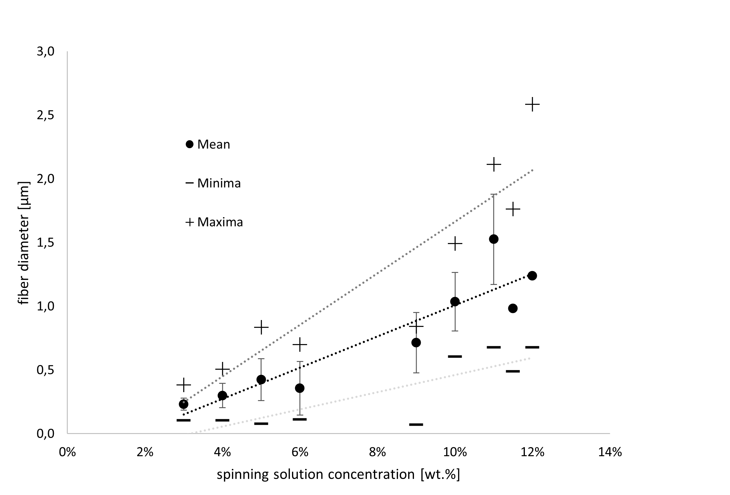
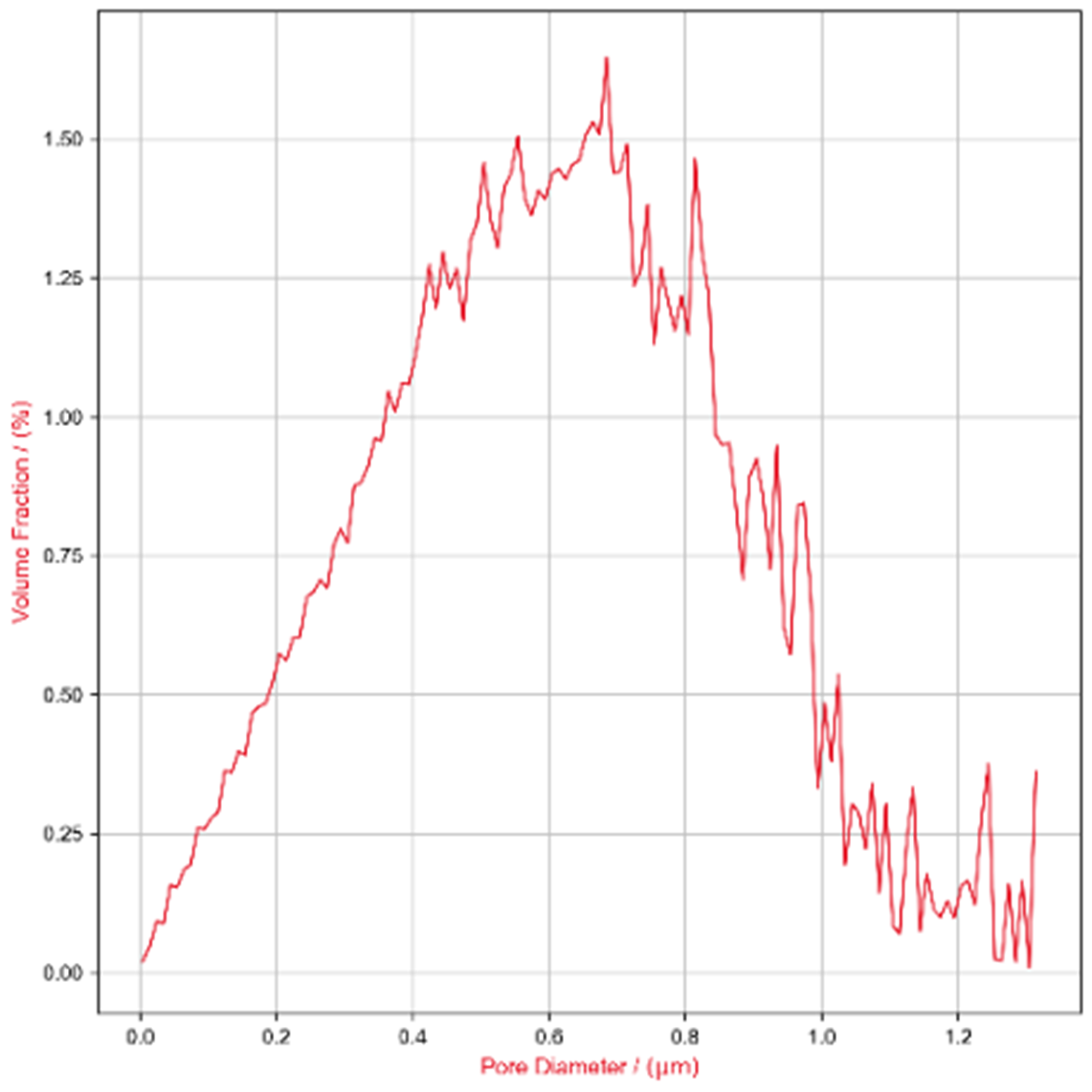
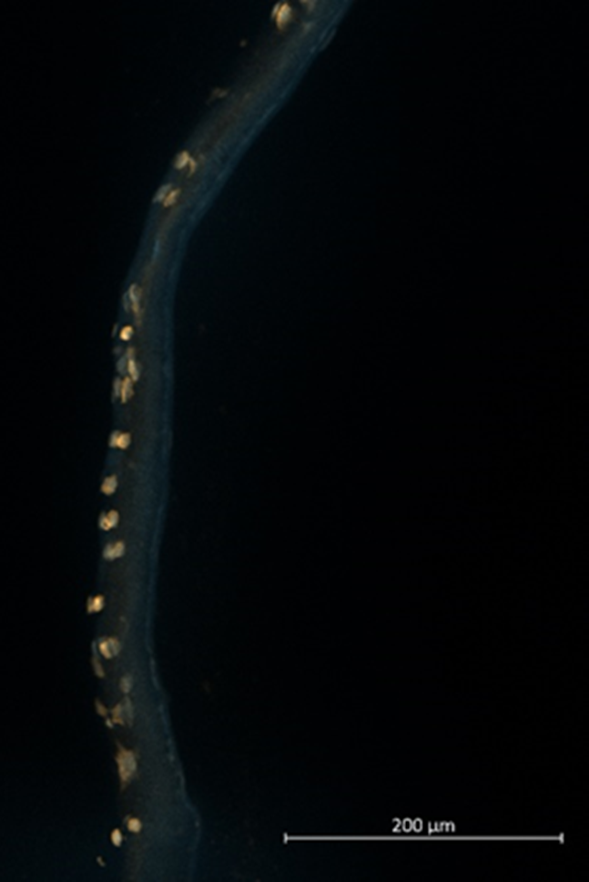
Barrier layer: To create submicroscale pores, the interactions between process and spinning solution parameters as well as fiber diameters were first investigated in detail. The spinning solution concentration (2–15%), the silk fibroin (SF)–polycaprolactone (PCL) ratio (0–100% SF), the influence of salt additives (up to 5 wt-% potassium chloride - KCl), the flow rate (0.5–2 ml/h), and the electric field strength (0–1.5 kV/cm) were systematically varied. It was found that fiber diameters can be specifically reduced, in particular by lowering the flow rate and the spinning solution concentration (Figure 4A) as well as by adding up to 66% SF. Higher SF content and salt additives lead to inhomogeneities in the resulting fiber diameters and shapes, as well as, in some cases, to embrittlement of the membranes. Varying the electric field strength (0–40 kV) also affected the fiber diameters, though only to a very small extent (approx. ± 22 nm); therefore, the preferred solution was selected here based on differences in handling during membrane production. Concentrations below 3% were not spinnable or resulted in irregular strand breaks. Based on these findings, the case group SF:PCL_1:2_3% was selected as the preferred solution. This case group exhibited nearly normally distributed pore sizes with a mean of 613 nm (Figure 4B). Approximately 6% of the pores had a diameter below the target value of 200 nm. To investigate migration behavior, the membranes were mounted in cell culture inserts (Cell-Crown™ inserts) and seeded with gingival epithelial cells for 72 h. After cultivation, cryosections were prepared, and the cytoskeleton and cell nuclei were stained. The barrier function was demonstrated over a period of 72 h (Figure 4C).
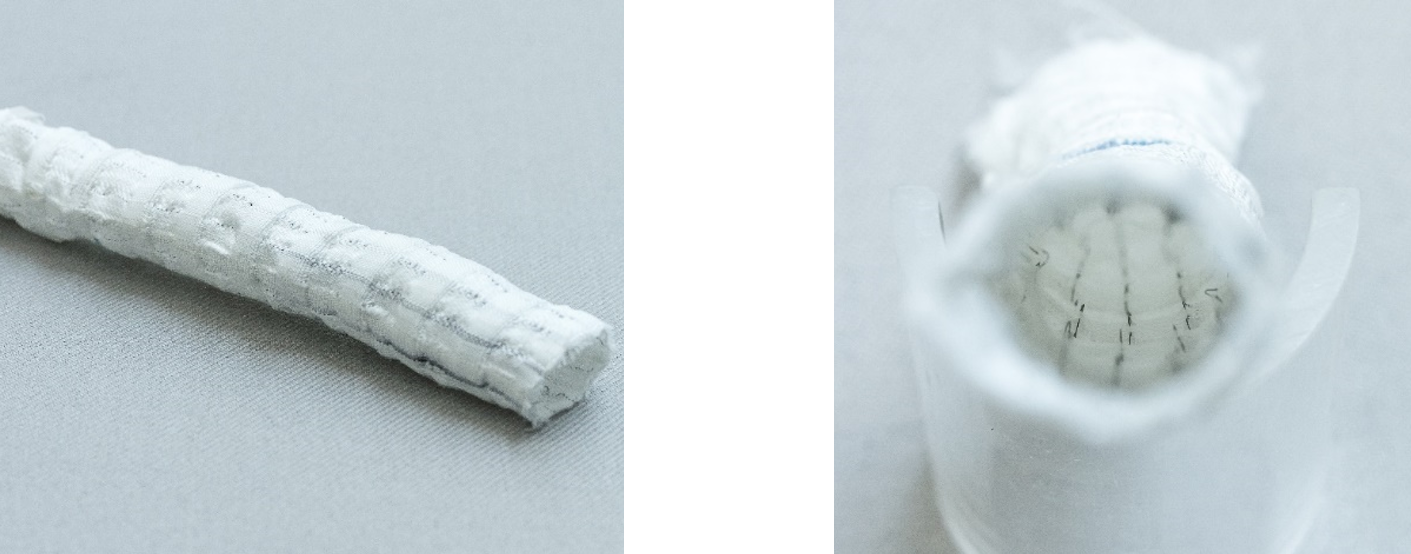
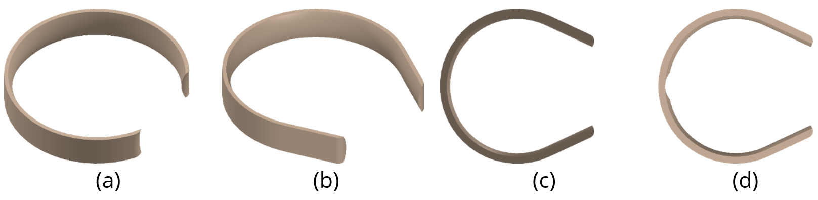
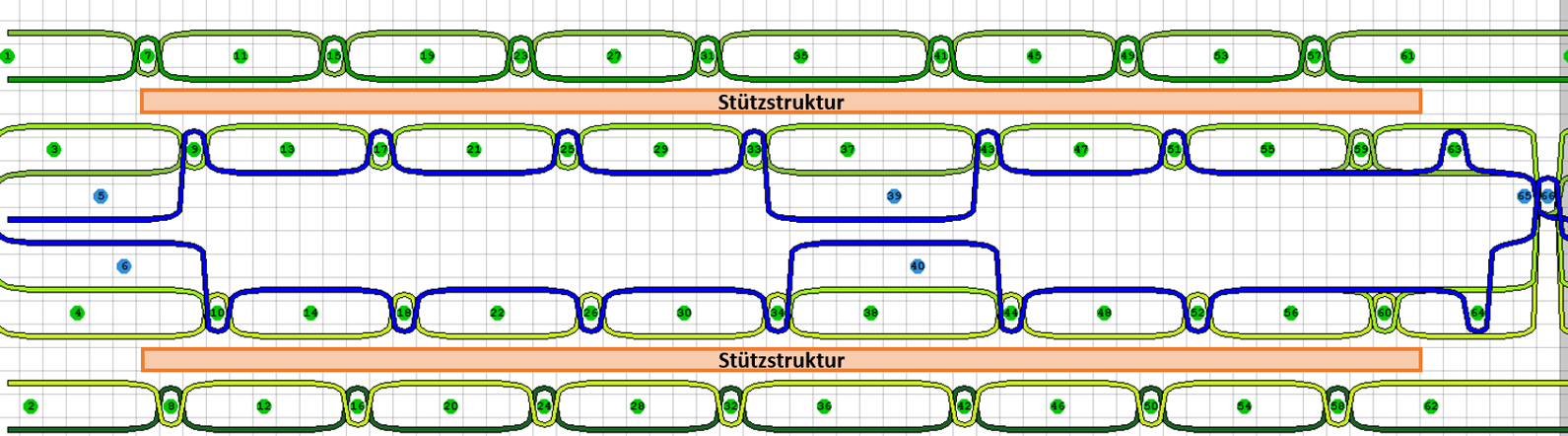
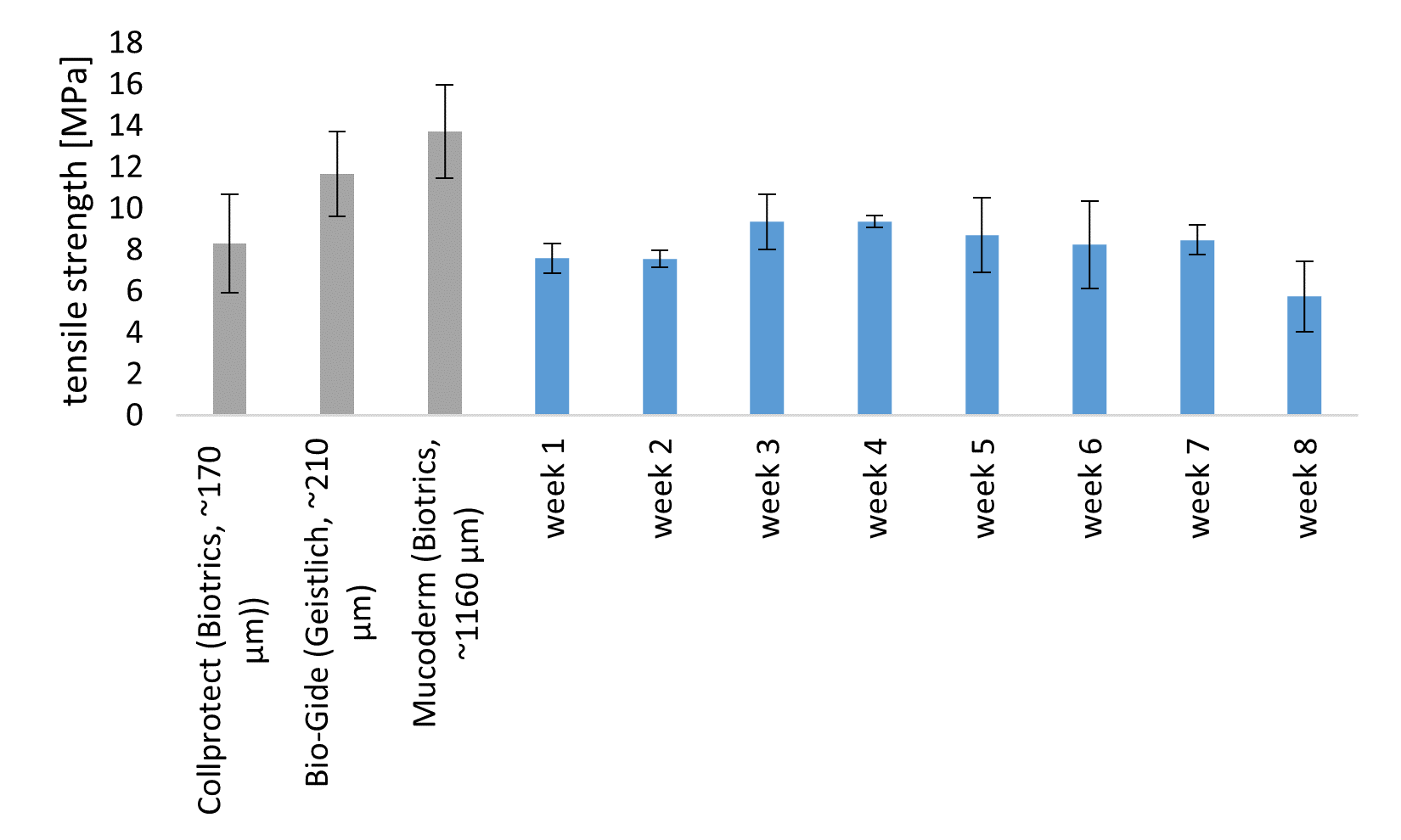
Mineral Layer: To achieve the desired structure of macroporous reinforcement structures, a tricalcium phosphate (TCP)-containing printing paste based on PCL was developed for the FAM. Mineral content of up to 50% was achieved without compromising printability. Using the FAM, constructs with triangular pore geometries were produced, as these offered advantages in terms of surgical handling, particularly with regard to flexibility. The 3D-printed specimens were subjected to enzymatic-catalyzed degradation over an 8-week period and, depending on the degree of degradation, were mechanically characterized in a humid environment and compared with commercially available, non-degraded GBR membranes (BioGuide, CollProtect, Mucoderm). To this end, four test specimens per week were incubated for 8 weeks in a degradation solution (physiological phosphate-buffered saline (PBS) and an enzyme cocktail consisting of protease XIV and lipase). The medium was changed twice each week to prevent the degradation solution from becoming saturated with degradation products. The enzyme concentration was set at 2.5 U/L per enzyme so that both the lipase and protease, considered individually, would reflect the total enzyme concentration in human saliva [9]. This increased enzyme concentration enables rapid degradation while also providing reliable data on the minimum shelf life of the developed GBR membrane in the human oral cavity. Initially, a slight increase in mass and tensile strength in the wet state was observed, which is presumably due to the deposition of salts on the surface (Figure 5). Starting in week 3, a continuous decline in mass and mechanical properties can be observed, although this decline is comparatively minor. Even after 7 weeks, the tensile strength remains in the range of 8 MPa and is thus comparable to the initial strength prior to degradation. Thus, the space-maintaining capacity of the developed GBR membrane was demonstrated for approximately 2 months, which is sufficient for the remodeling of natural bone tissue. The target value of > 5 MPa in a moist environment was maintained throughout the entire degradation period. Furthermore, a comparison was conducted with non-degrading, commercially available GBR membranes, demonstrating the comparability of the developed membrane. In addition, continuous calcium release was detected over the course of the degradation period, which may have a positive effect on bone regeneration.
To evaluate the osteoconductive effect of the mineral layer, the differentiation of bone progenitor cells (mesenchymal stem cells, MSCs) into osteogenic cells was investigated. Based on the staining of calcium deposits in the mineralized tissue (Alizarin Red S staining) and the quantitative analysis of osteogenic marker expression (qPCR of collagen I (Col1), alkaline phosphatase (ALP), osteocalcin (BGLAP), and osteopontin (SPP1)), a positive effect of the membrane developed in the project on bone tissue regeneration was demonstrated. Osteogenic induction was achieved by adding dexamethasone, β-glycerophosphate, and ascorbic acid for 28 days. Differentiation was controlled using MSCs without any material influence.
Summary: As a result of the OsteoMe project, three membrane layers were developed, each of which fulfills essential aspects of the requirements profile for GBR membranes in dentistry. Depending on the acidity of the chitosan as well as the contact time and pressure, the mucoadhesive layer develops adhesive forces in the range of 1 to 4 N, which are comparable to those of established fibrin glues. This enables the membrane to be applied to the defect site in a position-stable manner without the need for additional sutures or pins. The barrier layer is designed with open pores, allowing for the exchange of nutrients and signaling molecules, which can positively influence regeneration. At the same time, it was shown that the pore size is small enough to act as a barrier to rapidly proliferating oral mucosal cells, thereby providing the slow-growing bone tissue cells with sufficient time for regeneration. The 3D-printed mineral layer exhibits sufficiently high mechanical properties in simulated saliva to ensure a space-maintaining function. The tensile strengths achieved are comparable to those of commercially available GBR membranes and show virtually no decline during enzymatic-catalyzed degradation over eight weeks. Furthermore, it was demonstrated that the mineral layer exhibits osteoinductive properties and can thus actively support bone regeneration. Finally, methods were developed to ensure a delamination-free bonding of the individual layers. This allows the layers to be flexibly combined with one another, thereby expanding the range of possible applications.
Acknowledgments: The IGF project 01IF22810N of the Forschungskuratorium Textil e.V. research association was funded by the Federal Ministry for Economic Affairs and Energy through the DLR Project Management Agency as part of the Program for the Promotion of Industrial Collaborative Research (IGF), pursuant to a resolution of the German Bundestag.
Quellenverzeichnis:
[1] CHOLMAKOW-BODECHTEL, Constanze: Fünfte Deutsche Mundgesundheitsstudie (DMS V). JORDAN, Andreas Rainer (Hrsg.); MICHEELIS, Wolfgang (Hrsg.). Köln : Deutscher Zahnärzte Verlag DÄV, 2016
[2] CHA, Hyun-Suk ; KIM, Ji-Wan ; HWANG, Jong-Hyun ; AHN, Kang-Min: Frequency of bone graft in implant surgery. In: Maxillofacial plastic and reconstructive surgery 38 (2016), Nr. 1, S. 19
[3] MOY, Peter K. ; AGHALOO, Tara: Risk factors in bone augmentation procedures. In: Periodon-tology 2000 81 (2019), Nr. 1, S. 76–90
[4] KNÖFLER, Wolfram ; BARTH, Thomas ; GRAUL, Reinhard ; KRAMPE, Dietmar: Retrospective analysis of 10,000 implants from insertion up to 20 years-analysis of implantations using aug-mentative procedures. In: International journal of implant dentistry 2 (2016), Nr. 1, S. 25
[5] CABALLÉ-SERRANO, Jordi ; MUNAR-FRAU, Antonio ; ORTIZ-PUIGPELAT, Octavi ; SOTO-PENALOZA, David ; PEÑARROCHA, Miguel ; HERNÁNDEZ-ALFARO, Federico: On the search of the ideal barrier membrane for guided bone regeneration. In: Journal of clinical and experi-mental dentistry 10 (2018), Nr. 5, e477-e483
[6] JIMÉNEZ GARCIA, J. ; BERGHEZAN, S. ; CARAMÊS, J. M. M. ; DARD, M. M. ; MARQUES, D. N. S.: Effect of cross-linked vs non-cross-linked collagen membranes on bone: A systematic re-view. In: Journal of periodontal research 52 (2017), Nr. 6, S. 955–964
[7] BUTENSCHÖN, Sina: Prävalenz periimplantärer Entzündungen bei teilbezahnten Patienten nach einer minimalen Beobachtungsdauer von 10 Jahren - eine retrospektive Querschnittsstu-die. Göttingen, Georg-August-Universität zu Göttingen. Dissertation. 2019
[8] RAKIC, Mia ; GALINDO-MORENO, Pablo ; MONJE, Alberto ; RADOVANOVIC, Sandro ; WANG, Hom-Lay ; COCHRAN, David ; SCULEAN, Anton ; CANULLO, Luigi: How frequent does peri-implantitis occur? A systematic review and meta-analysis. In: Clinical oral investigations 22 (2018), Nr. 4, S. 1805–1816
[9] CHAUNCEY, Howard Haskell. The chemical composition of human sal
Contact: lukas.benecke@tu-dresden.de
Technische Universität Dresden
Fakultät Maschinenwesen
Institut für Textilmaschinen und Textile Hochleistungswerkstofftechnik (ITM)
01062 Dresden
FILK Freiberg Institute gGmbH
Meißner Ring 1-5
09599 Freiberg
Deutschland
https://www.filkfreiberg.de/