Artificial Muscle Fibers Could Serve as Cell Scaffolds
In two new studies, North Carolina State University researchers designed and tested a series of textile fibers that can change shape and generate force like a muscle. In the first study, the researchers focused on the materials’ influence on the artificial muscles’ strength and contraction length. The findings could help researchers tailor the fibers for different applications.
In the second, proof-of-concept study, the researchers tested their fibers as scaffolds for live cells. Their findings suggest the fibers – known as “fiber robots” – could potentially be used to develop 3D models of living, moving systems in the human body.
“We found that our fiber robot is a very suitable scaffold for the cells, and we can alter the frequency and contraction ratio to create a more suitable environment for cells,” said Muh Amdadul Hoque, graduate student in textile engineering, chemistry and science at NC State. “These were proof-of concept studies; ultimately, our goal is to see if we can study these fibers as a scaffold for stem cells, or use them to develop artificial organs in future studies.”
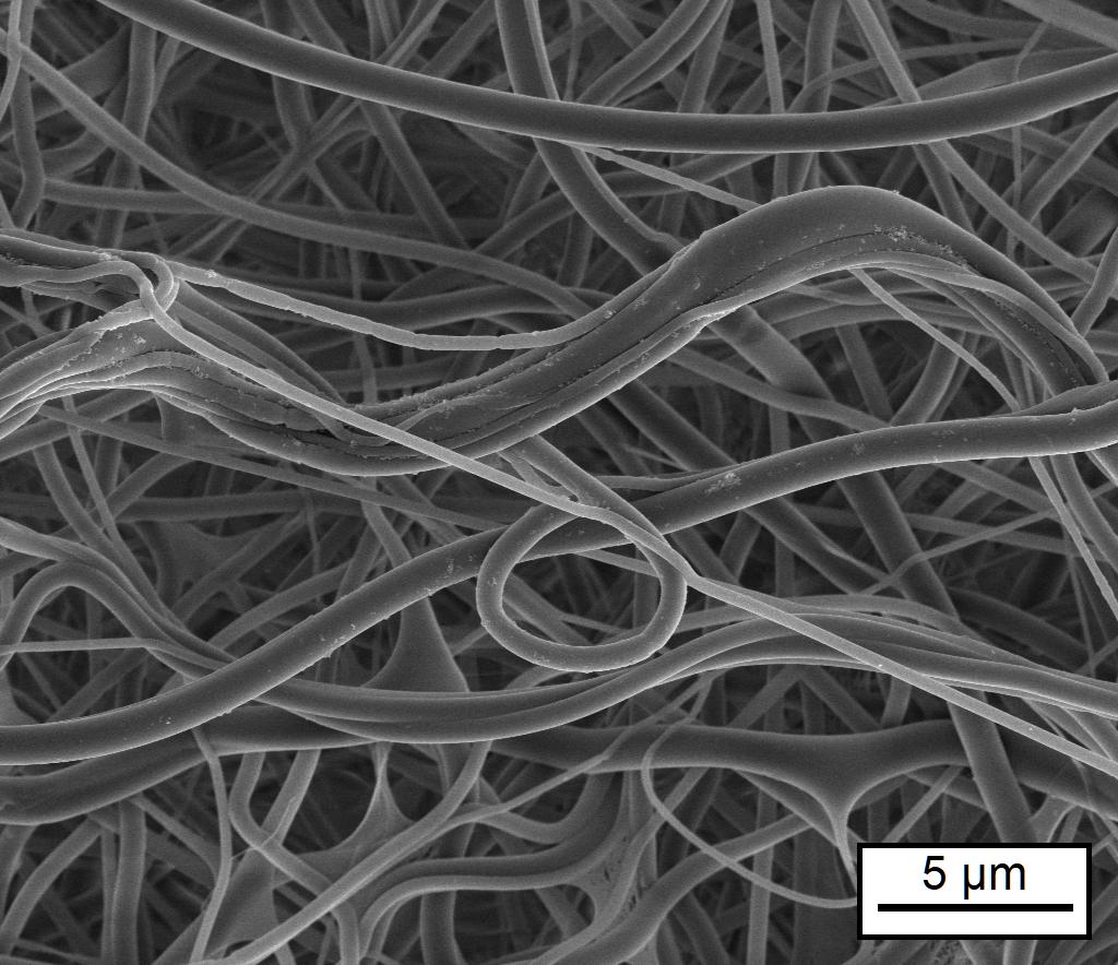
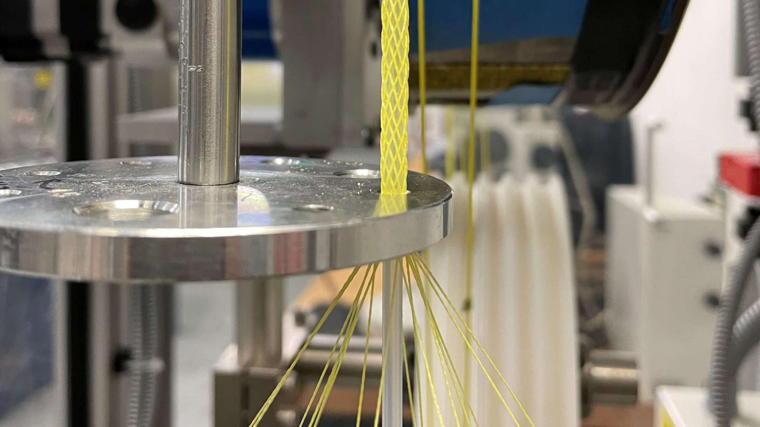
Researchers made the shape-changing fibers by encapsulating a balloon-like tube, made of a material similar to rubber, in a braided textile sheath. Inflating the interior balloon with an air pump makes the braided sheath expand, causing it to shorten.
The researchers measured the force and contraction rates of fibers made from different materials in order to understand the relationship between material and performance. They found that stronger, larger diameter yarns generated a stronger contraction force. In addition, they found that the material used to make the balloon impacted the magnitude of the contraction and generated force.
“We found that we could tailor the material properties to the required performance of the device,” said Xiaomeng Fang, assistant professor of textile engineering, chemistry and science at NC State. “We also found that we can make this device small enough so we can potentially use it in fabric formation and other textile applications, including in wearables and assistive devices.”
In a follow-up study, researchers evaluated whether they could use the shape-changing fibers as a scaffold for fibroblasts, a cell type found in connective tissues that help support other tissues or organs.
“The idea with stretching is to mimic the dynamic nature of how your body moves,” said Jessica Gluck, assistant professor of textile engineering, chemistry and science at NC State, and a study co-author.
They studied the cells’ response to the motion of the shape-changing fibers, and to different materials used in the fibers’ construction. They found the cells were able to cover and even penetrate the fiber robot’s braiding sheath. However, they saw decreases in the cells’ metabolic activity when the fiber robot’s contraction extended beyond a certain level, compared to a device made of the same material that they kept stationary.
The researchers are interested in building on the findings to see if they could use the fibers as a 3D biological model, and to investigate whether movement would impact cell differentiation. They said their model would be an advance over other existing experimental models that have been developed to show cellular response to stretching and other motion, since they can only move in two dimensions.
“Typically, if you want to add stretch or strain on cells, you would put them onto a plastic dish, and stretch them in one or two directions,” Gluck said. “In this study, we were able to show that in this 3D dynamic culture, the cells can survive for up to 72 hours.
“This is particularly useful for stem cells,” Gluck added. “What we could do in the future is look at what could happen at the cellular level with mechanical stress on the cells. You could look at muscle cells and see how they’re developing, or see how the mechanical action would help differentiate the cells.”
The study, “Effect of Material Properties on Fiber-Shaped Pneumatic Actuators Performance” was published in Actuators on March 18. Emily Petersen was a co-author. The study was funded by start-up funding awarded to Fang from the Department of Textile Engineering, Chemistry and Science at NC State.
The study, “Development of a Pneumatic-Driven Fiber-Shaped Robot Scaffold for Use as a Complex 3D Dynamic Culture System” was published online in Biomimetics on April 21. In addition to Gluck, Hoque and Fang, co-authors included Nasif Mahmood, Kiran M. Ali, Eelya Sefat, Yihan Huang, Emily Petersen and Shane Harrington. The study was funded by the NC State Wilson College of Textiles, the Department of Textile Engineering, Chemistry and Science and the Wilson College of Textiles Research Opportunity Seed Fund Program.
North Carolina State University, Laura Oleniacz. Übersetzung Textination