Textination Newsline

Reset
8 results
Empa researcher Simon Annaheim is working to develop a mattress for newborn babies. Image: Empa
11.03.2024

Medical textiles and sensors: Smart protection for delicate skin

Skin injuries caused by prolonged pressure often occur in people who are unable to change their position independently – such as sick newborns in hospitals or elderly people. Thanks to successful partnerships with industry and research, Empa scientists are now launching two smart solutions for pressure sores.

If too much pressure is applied to our skin over a long period of time, it becomes damaged. Populations at high risk of such pressure injuries include people in wheelchairs, newborns in intensive care units and the elderly. The consequences are wounds, infections and pain.

Treatment is complex and expensive: Healthcare costs of around 300 million Swiss francs are incurred every year. "In addition, existing illnesses can be exacerbated by such pressure injuries," says Empa researcher Simon Annaheim from the Biomimetic Membranes and Textiles laboratory in St. Gallen. According to Annaheim, it would be more sustainable to prevent tissue damage from occurring in the first place. Two current research projects involving Empa researchers are now advancing solutions: A pressure-equalizing mattress for newborns in intensive care units and a textile sensor system for paraplegics and bedridden people are being developed.

Optimally nestled at the start of life
The demands of our skin are completely different depending on age: In adults, the friction of the skin on the lying surface, physical shear forces in the tissue and the lack of breathability of textiles are the main risk factors. In contrast, the skin of newborns receiving intensive care is extremely sensitive per se, and any loss of fluid and heat through the skin can become a problem. "While these particularly vulnerable babies are being nursed back to health, the lying situation should not cause any additional complications," says Annaheim. He thinks conventional mattresses are not appropriate for newborns with very different weights and various illnesses. Annaheim's team is therefore working with researchers from ETH Zurich, the Zurich University of Applied Sciences (ZHAW) and the University Children's Hospital Zurich to find an optimal lying surface for babies' delicate skin. This mattress should be able to adapt individually to the body in order to help children with a difficult start in life.

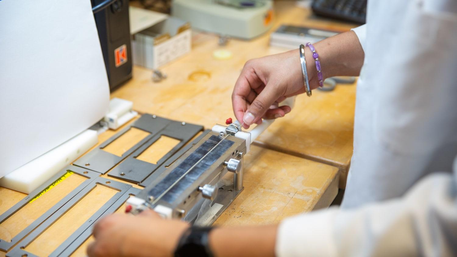
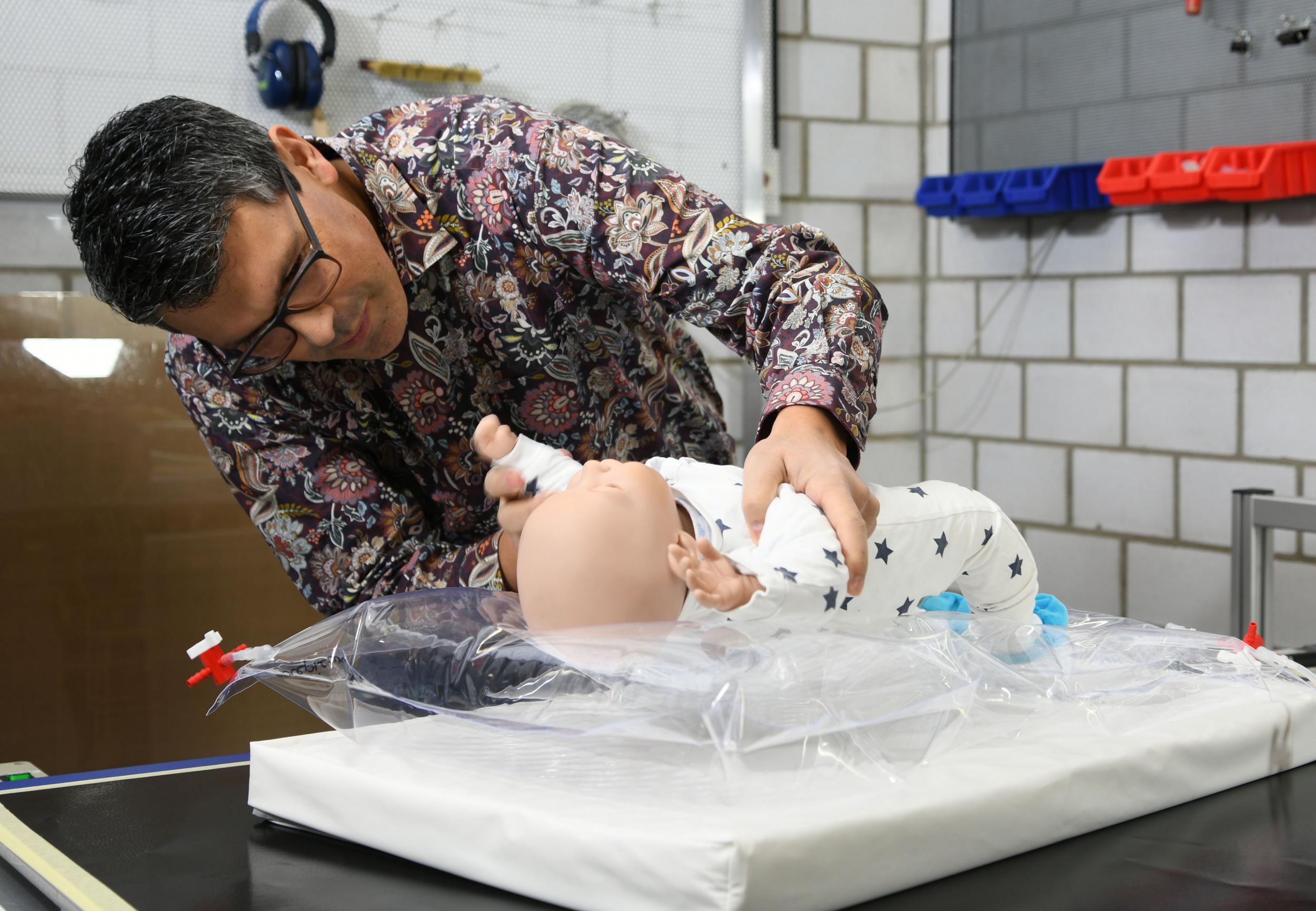
In order to do this, the researchers first determined the pressure conditions in the various regions of the newborn's body. "Our pressure sensors showed that the head, shoulders and lower spine are the areas with the greatest risk of pressure sores," says Annaheim. These findings were incorporated into the development of a special kind of air-filled mattress: With the help of pressure sensors and a microprocessor, its three chambers can be filled precisely via an electronic pump so that the pressure in the respective areas is minimized. An infrared laser process developed at Empa made it possible to produce the mattress from a flexible, multi-layered polymer membrane that is gentle on the skin and has no irritating seams.

After a multi-stage development process in the laboratory, the first small patients were allowed to lie on the prototype mattress. The effect was immediately noticeable when the researchers filled the mattress with air to varying degrees depending on the individual needs of the babies: Compared to a conventional foam mattress, the prototype reduced the pressure on the vulnerable parts of the body by up to 40 percent.

Following this successful pilot study, the prototype is now being optimized in the Empa labs. Simon Annaheim and doctoral student Tino Jucker will soon be starting a larger-scale study with the new mattress with the Department of Intensive Care Medicine & Neonatology at University Children's Hospital Zurich.

Intelligent sensors prevent injuries
In another project, Empa researchers are working on preventing so-called pressure ulcer tissue damage in adults. This involves converting the risk factors of pressure and circulatory disorders into helpful warning signals.

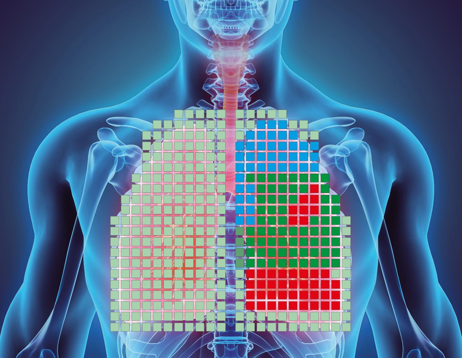
If you lie in the same position for a long time, pressure and circulatory problems lead to an undersupply of oxygen to the tissue. While the lack of oxygen triggers a reflex to move in healthy people, this neurological feedback loop can be disrupted in people with paraplegia or coma patients, for example. Here, smart sensors can help to provide early warning of the risk of tissue damage.

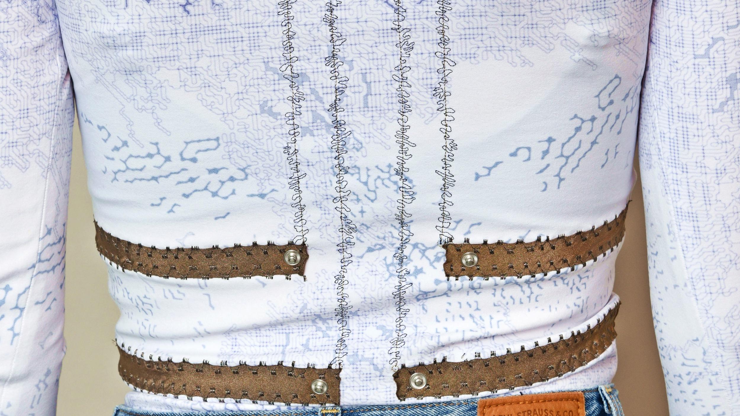
In the ProTex project, a team of researchers from Empa, the University of Bern, the OST University of Applied Sciences and Bischoff Textil AG in St. Gallen has developed a sensor system made of smart textiles with associated data analysis in real time. "The skin-compatible textile sensors contain two different functional polymer fibers," says Luciano Boesel from Empa's Biomimetic Membranes and Textiles laboratory in St. Gallen. In addition to pressure-sensitive fibers, the researchers integrated light-conducting polymer fibers (POFs), which are used to measure oxygen. "As soon as the oxygen content in the skin drops, the highly sensitive sensor system signals an increasing risk of tissue damage," explains Boesel. The data is then transmitted directly to the patient or to the nursing staff. This means, for instance, that a lying person can be repositioned in good time before the tissue is damaged.

Patented technology
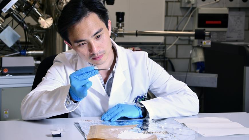
The technology behind this also includes a novel microfluidic wet spinning process developed at Empa for the production of POFs. It allows precise control of the polymer components in the micrometer range and smoother, more environmentally friendly processing of the fibers. The microfluidic process is one of three patents that have emerged from the ProTex project to date.

Another product is a breathable textile sensor that is worn directly on the skin. The spin-off Sensawear in Bern, which emerged from the project in 2023, is currently pushing ahead with the market launch. Empa researcher Boesel is also convinced: "The findings and technologies from ProTex will enable further applications in the field of wearable sensor technology and smart clothing in the future."

Source:

Dr. Andrea Six, Empa

Wearable Robots for Parkinson’s Disease Image: Tom Claes, unsplash
19.02.2024

Wearable Robots for Parkinson’s Disease

Freezing is one of the most common and debilitating symptoms of Parkinson’s disease, a neurodegenerative disorder that affects more than 9 million people worldwide. When individuals with Parkinson’s disease freeze, they suddenly lose the ability to move their feet, often mid-stride, resulting in a series of staccato stutter steps that get shorter until the person stops altogether. These episodes are one of the biggest contributors to falls among people living with Parkinson’s disease.

Today, freezing is treated with a range of pharmacological, surgical or behavioral therapies, none of which are particularly effective. What if there was a way to stop freezing altogether?

Researchers from the Harvard John A. Paulson School of Engineering and Applied Sciences (SEAS) and the Boston University Sargent College of Health & Rehabilitation Sciences have used a soft, wearable robot to help a person living with Parkinson’s walk without freezing. The robotic garment, worn around the hips and thighs, gives a gentle push to the hips as the leg swings, helping the patient achieve a longer stride.

The device completely eliminated the participant’s freezing while walking indoors, allowing them to walk faster and further than they could without the garment’s help.

“We found that just a small amount of mechanical assistance from our soft robotic apparel delivered instan-taneous effects and consistently improved walking across a range of conditions for the individual in our study,” said Conor Walsh, the Paul A. Maeder Professor of Engineering and Applied Sciences at SEAS and co-corresponding author of the study.

The research demonstrates the potential of soft robotics to treat this frustrating and potentially dangerous symptom of Parkinson’s disease and could allow people living with the disease to regain not only their mobility but their independence.

For over a decade, Walsh’s Biodesign Lab at SEAS has been developing assistive and rehabilitative robotic technologies to improve mobility for individuals’ post-stroke and those living with ALS or other diseases that impact mobility. Some of that technology, specifically an exosuit for post-stroke gait retraining, received support from the Wyss Institute for Biologically Inspired Engineering, and Harvard’s Office of Technology Development coordinated a license agreement with ReWalk Robotics to commercialize the technology.

In 2022, SEAS and Sargent College received a grant from the Massachusetts Technology Collaborative to support the development and translation of next-generation robotics and wearable technologies. The research is centered at the Move Lab, whose mission is to support advances in human performance enhancement with the collaborative space, funding, R&D infrastructure, and experience necessary to turn promising research into mature technologies that can be translated through collaboration with industry partners. This research emerged from that partnership.

“Leveraging soft wearable robots to prevent freezing of gait in patients with Parkinson’s required a collaboration between engineers, rehabilitation scientists, physical therapists, biomechanists and apparel designers,” said Walsh, whose team collaborated closely with that of Terry Ellis,  Professor and Physical Therapy Department Chair and Director of the Center for Neurorehabilitation at Boston University.

Leveraging soft wearable robots to prevent freezing of gait in patients with Parkinson’s required a collaboration between engineers, rehabilitation scientists, physical therapists, biomechanists and apparel designers.

The team spent six months working with a 73-year-old man with Parkinson’s disease, who — despite using both surgical and pharmacologic treatments — endured substantial and incapacitating freezing episodes more than 10 times a day, causing him to fall frequently. These episodes prevented him from walking around his community and forced him to rely on a scooter to get around outside.

In previous research, Walsh and his team leveraged human-in-the-loop optimization to demonstrate that a soft, wearable device could be used to augment hip flexion and assist in swinging the leg forward to provide an efficient approach to reduce energy expenditure during walking in healthy individuals.

Here, the researchers used the same approach but to address freezing. The wearable device uses cable-driven actuators and sensors worn around the waist and thighs. Using motion data collected by the sensors, algorithms estimate the phase of the gait and generate assistive forces in tandem with muscle movement.

The effect was instantaneous. Without any special training, the patient was able to walk without any freezing indoors and with only occasional episodes outdoors. He was also able to walk and talk without freezing, a rarity without the device.

“Our team was really excited to see the impact of the technology on the participant’s walking,” said Jinsoo Kim, former PhD student at SEAS and co-lead author on the study.

During the study visits, the participant told researchers: “The suit helps me take longer steps and when it is not active, I notice I drag my feet much more. It has really helped me, and I feel it is a positive step forward. It could help me to walk longer and maintain the quality of my life.”

“Our study participants who volunteer their time are real partners,” said Walsh. “Because mobility is difficult, it was a real challenge for this individual to even come into the lab, but we benefited so much from his perspective and feedback.”

The device could also be used to better understand the mechanisms of gait freezing, which is poorly understood.

“Because we don’t really understand freezing, we don’t really know why this approach works so well,” said Ellis. “But this work suggests the potential benefits of a ’bottom-up’ rather than ’top-down’ solution to treating gait freezing. We see that restoring almost-normal biomechanics alters the peripheral dynamics of gait and may influence the central processing of gait control.”

The research was co-authored by Jinsoo Kim, Franchino Porciuncula, Hee Doo Yang, Nicholas Wendel, Teresa Baker and Andrew Chin. Asa Eckert-Erdheim and Dorothy Orzel also contributed to the design of the technology, as well as Ada Huang, and Sarah Sullivan managed the clinical research. It was supported by the National Science Foundation under grant CMMI-1925085; the National Institutes of Health under grant NIH U01 TR002775; and the Massachusetts Technology Collaborative, Collaborative Research and Development Matching Grant.

Source:

The research is published in Nature Medicine.
Source Leah Burrows
Harvard John A. Paulson. School of Engineering and Applied Sciences

Better Manufacturing Method for Wound Closures (c) Wilson College of Textiles
03.01.2024

Better Manufacturing Method for Wound Closures

If you’ve ever gotten stitches or had surgery, you may have had a suture. They’re the threads used to close wounds or join tissues together for other purposes.

But did you know that there are different types of sutures which can have an effect on your experience at the doctor or surgeon’s office?

Barbed sutures, for example, can reduce the amount of time you spend on the operating table and lower the likelihood of surgical complications. That type of suture has its roots in the Triangle and is being advanced by students and faculty at the Wilson College of Textiles.

Dr. Gregory Ruff, a nationally-renowned plastic surgeon, first invented the innovative closure in 1991, just down the road in Chapel Hill, North Carolina.

“I was thinking about the fact that we sew wounds together with a loop and a knot and if you tie it too tight, it can constrict the circulation and kill the tissue in that loop,” Dr. Ruff remembers.

“I was thinking about animals, and a porcupine’s quill came to mind. And the aha moment was, ‘What if we put a quill on one side of the wound and another one on the other side of the wound, so there’s no loop: the barbs go in but they don’t come out?’”

As the name suggests, barbed sutures have small projections shooting out of them that can latch onto tissues: think about barbed wire or a fishing hook. Those “quills,” or barbs, allow the suture to self-anchor. Since no knot is needed to secure the suture, the closure is faster, and the lack of knots and constricting loops promotes healing. This also allows surgeons to schedule more surgeries.

Soon after his aha moment, Dr. Ruff started his own company, Quill Medical, to fabricate these barbed sutures. While he had the medical expertise and a solid business partner, Dr. Ruff was looking for someone who could advise him in terms of the material makeup of the suture. The Wilson College’s Biomedical Textile Research Group, under the direction of Professor Martin King, quickly proved to be the perfect partner.

Using the Wilson College’s labs, King’s graduate students conducted a number of tests on Ruff’s sutures across different types of tissues (such as skin, muscle, etc.). One of those students, Nilesh Ingle, found that the barbed sutures worked best when the angles of the barbs were tailored specifically to the type of tissue being sutured.

Years later, one of King’s current graduate students is building on that research insight.
 
Understanding challenges and innovating solutions
Nearly three decades after the barbed suture’s invention, the majority of surgeons still use conventional sutures despite the advantages documented by researchers and surgeons. Why?

Karuna Nambi Gowri, a fiber and polymer science doctoral student in King’s research group, says it comes down to two reasons. The first of these is resistance to change. Most practicing surgeons learned how to use a suture before barbed sutures became more broadly available.

The second obstacle to the use of barbed sutures is procuring them. Barbed sutures tend to be both expensive and low in supply. That’s because the current process for making them (mechanical and blade-based) is inefficient in terms of both time and resources.

That’s where Nambi Gowri’s research with the Wilson College’s Biomedical Textiles Research Group comes in. She’s developing a faster and cheaper method for making the same quality of barbed suture.

“If I fabricate using a laser, the fabrication time is pretty short compared to a mechanical barbing technique,” Nambi Gowri says.

Moving from a mechanical method to a laser method has another advantage.

“The manipulation of the barbed suture itself is easier using a laser,” she says.

In other words, using the lasers will allow Nambi Gowri to apply the custom barb geometries, or angles, suggested by prior researchers on a commercial scale. These custom geometries will allow the barbed suture to be optimized for the type of tissue it will be connecting.

In addition to the new process, Nambi Gowri is also developing a new suture.

“I’m the first one to actually study Catgut barbed sutures,” she explains.

Catgut was actually one of the earliest materials used to make sutures. The filament is made from tissue taken from an animal’s stomach – especially cattle stomachs – hence the name. While the industry had moved away from this material in favor of synthetic polymers, Nambi Gowri sees the potential for Catgut in barbed sutures because of their quick degradation rate.

“These are useful external wound closures,” she says. “Because our body contains so much collagen and Catgut is made up of 90% collagen, it’s a more suitable polymer that can be used in human tissue.”

Hands-on experience informs research
In the meantime, Nambi Gowri has gained hands-on experience to inform her research by fabricating all of the barbed sutures used in Dr. Ruff’s micro facelift surgeries.

The surgery itself is made possible because of the shape and the material composition of the sutures: poly 4-hydroxybutyrate (P4HB). This polymer is already present naturally within our bodies, so sutures made from P4HB are naturally and safely absorbed by the body over time. That means patients don’t have to schedule an appointment after surgery for the sutures to be removed.
 
P4HB also provides the perfect combination of strength and elasticity to hold up the facial tissue until the wound has healed. The barbs, on the other hand, allow for the suture to be placed and stay secure within the skin without the need for large incisions.

“That skin tightens up right away,” Dr. Ruff says of the procedure, which draws patients from across the country. “So I don’t have to remove hair, and I don’t have to put a scar at the hairline.”

“These sutures are not available commercially anywhere in the world. So, to be able to mechanically barb different size sutures in a reliable and consistent manner for use in clinical practice, requires skill, experience and knowledge of quality control,” Professor King says of Nambi Gowri’s work.

This has given Karuna a hands-on understanding of the sutures she’s hoping to improve upon.

She says her fiber and polymer science knowledge has played a key role in helping her approach all sides of her research.

“All the analytical characterization techniques that are used for characterization of sutures – like identifying mechanical properties and measuring tensile strength – is actually from my knowledge of textiles,” she says. “I’m applying my polymer chemistry knowledge  to make sure that the laser doesn’t cause the sutures to degrade, melt or experience thermal damage.”

What’s next?
As she works to patent her designs, Nambi Gowri feels confident that her dissertation will set her up for success in the research and development (R&D) field after graduation.

In the meantime, she’s already finding out about the ways her research can have a broader impact.

“Dr. Dan Duffy, DVM, a surgeon at the NC State College of Veterinary Medicine is also interested in using barbed sutures to repair torn and failed tendons on his animals, but he finds the cost of buying commercial barbed sutures prohibitively expensive. So we need to collaborate,” King says. “Karuna to the rescue!”

Source:

North Carolina State University, Sarah Stone

Silk Provides the Building Blocks to Transform Modern Medicine Photo: Jenna Schad
31.10.2023

Silk Provides the Building Blocks to Transform Modern Medicine

Tufts researchers harness protein from silk to make virus-sensing gloves, surgical screws that dissolve in your body, and other next-generation biomedical materials

About a mile northwest of Tufts’ Medford/Somerville campus, on the fourth floor of a refurbished woolen factory, there is a shrine to silk. Glass vases filled with silkworm cocoons and washed silk fibers sit artfully on a shelf across from a colorful drawing of the life cycle of Bombyx mori, the domesticated silk moth. Farther in, more cocoons in wall-mounted cases border a large, close-up image of silk fibers, and displays hold dozens of prototypes made from silk, including smart fabrics, biosensors, a helmet that changes color upon impact, and potential replacements for materials like leather, plastic, and particle board.

The only things missing are the silkworms themselves, but Fiorenzo Omenetto, the director of Silklab and the Frank C. Doble Professor of Engineering at Tufts, said they will be arriving soon. The lab is building a terrarium so that visitors can view the animals.
“We’re going to have a celebration of silkworms and moths,” Omenetto said.

Silk has been cultivated and harvested for thousands of years. It is best known for the strong, shimmering fabric that can be woven from its fibers, but it also has a long history of use in medicine to dress injuries and suture wounds. At Silklab, Omenetto and his colleagues are building on silk’s legacy, proving that this ancient fiber could help create the next generation of biomedical materials.

Silk moth caterpillars, known as silkworms, extrude a single sticky strand of silk from their mouths to form cocoons, which are harvested by silk farmers to make silk thread. At its core, silk is a mixture of two proteins: fibroin, which provides the fiber’s structure, and sericin, which binds it together. With a few steps in the lab, Tufts researchers can remove the sericin and dissolve the fibers, turning a dry cocoon into a fibroin-filled liquid.

“Nature builds structural proteins that are very tough and very strong,” Omenetto said. “Your bricks are these fibroin proteins floating in water. From there, you can build whatever you want.”
Starting with shipments of dried cocoons from silk farms, Omenetto and his colleagues have been able to create gels, sponges, clear plastic-like sheets, printable inks, solids that look like amber, dippable coatings, and much more.

“Each of the materials that you make can contain all these different functions, and there’s only 24 hours in a day,” Omenetto said with a laugh. “This is why I don’t sleep.”

Biocompatible and Biodegradable
When Omenetto arrived at Tufts almost two decades ago, his research was focused on lasers and optics—silk wasn’t in the picture. But a chance conversation with David Kaplan, the Stern Family Professor of Engineering and chair of the biomedical engineering department, set him on a new path.

Kaplan, who has been working with silk since the early ’90s, was designing a silk scaffold that would help rebuild a person’s cornea, allowing cells to grow between the layers. He needed a way to ensure that the growing cells would have enough oxygen and showed the small, transparent sheet to Omenetto, who was immediately intrigued by the material. Omenetto was able to use his lab’s lasers to put tiny holes in Kaplan’s silk cornea. More collaborations quickly followed.
“We’ve worked together incessantly since then,” Kaplan said.

One of those lines of research has been finding ways to use silk to help repair and regrow bone, blood vessels, nerves, and other tissue. Silk is biocompatible, meaning it doesn’t cause harm in the body and breaks down in predictable ways. With the right preparation, silk materials can provide necessary strength and structure while the body is healing.

“You can mold and shape silk to whatever you need, and it will hold that volume while the native tissue regrows into the space and the silk material degrades,” Kaplan said. “Eventually it’s 100 percent gone, and you’re back to your normal tissue.”

Some of this work has already been approved for use by the U.S. Food and Drug Administration. A company called Sofregen, which spun out of Kaplan and Omenetto’s research, is using an injectable silk-based gel to repair damaged vocal cords, the tissues that regulate air flow and help us speak.

On their own, sturdy silk structures can keep their size, shape, and function for years before degrading. But in some instances, such as those involving surgical screws and plates intended for use in rapidly growing children, this pace would be too slow. The researchers had to find a way to speed up the time it takes for dense silk biomaterials to break down. They introduced an enzyme that our bodies produce naturally into the silk to hasten the breakdown process. The idea is that the enzyme would sit dry and inactive within the silk device until the structure is installed in a person, then the device would hydrate and activate the enzyme to digest the material more rapidly.

“We can titer in just the right amount of enzyme to make a screw go away in a week, a month, a year,” Kaplan said. “We have control over the process.”

Currently, Kaplan and his lab are working on other small, degradable medical devices that would help cut down on the number of surgeries that patients need. Ear tubes, for example, are often surgically implanted to help alleviate chronic ear infections and then need to be surgically removed. Kaplan and his colleagues have designed silk-based ear tubes that degrade on their own and can even carry antibiotics.

“As someone with a daughter who went through six surgeries on her ear, I know how helpful this could be,” Kaplan said.

Source:

Laura Castañón, Tufts University, Massachusetts USA

Functional textiles – an alternative to antibiotics University of Borås
04.07.2023

Functional textiles – an alternative to antibiotics

Tuser Biswas conducts research that aims to develop modern medical textiles that are good for both the environment and human health. Textiles with antimicrobial properties could reduce the use of antibiotics.

Tuser Biswas conducts research that aims to develop modern medical textiles that are good for both the environment and human health. Textiles with antimicrobial properties could reduce the use of antibiotics.

His work involves research and teaching activities within the area of textile material technology. The current research involves resource-efficient inkjet printing of functional materials on various textile surfaces for advanced applications.
 
The conventional textile industry devours natural resources in the form of water, energy, and chemicals. A more resource-efficient way to produce textiles is with ink jet printing. Tuser Biswas, who recently defended his doctoral thesis in Textile Material Technology, seeks to develop methods for functional textiles. He has shown that it is possible to print enzymes on textiles. These are proteins that function as catalysts in the body, as they set chemical processes in motion without themselves changing. They could, for example, be used in medical textiles with antimicrobial properties or to measure biological or chemical reactions.

“Ever since the industrial revolution, our society has used an abundance of synthetic and harsh chemicals. Our research works to replace these chemicals with environmentally friendly and bio-based materials,” said Tuser Biswas.
 
Promising results with enzymes on textiles
Developing a good enzyme ink was not entirely easy and it took a number of attempts before he finally, to his great joy, had successful results. Tuser Biswas explained that the most important result is to show how a printed enzyme could bind another enzyme to the surface of a fabric. Although the activity of the enzymes decreased by 20-30 percent after printing, the results are still promising for future applications. At the same time, the work has provided new knowledge about many fundamental questions about printing biomaterials on fabric.

“Before starting the project, we found several related studies that focused on producing a finished product. But we wanted to study the fundamental challenges of this subject, and now we know how to make it work,” said Tuser Biswas.

He is now seeking funding to continue researching the subject and has so far received a grant from the Sjuhärad Savings Bank Foundation. During the Days of Knowledge event in April 2023, he presented his research to representatives from the City of Borås and business, the Sjuhärad Savings Bank Foundation, and the University of Borås.
     
Medical textiles instead of antibiotics
Tuser Biswas hopes that continued research in textile technology can provide alternatives to using antibiotics. With increasing antibiotic resistance, it is an important issue not only locally but worldwide.

“Instead of treating the patient with a course of antibiotics, one can act preventively and more effectively by damaging the bacteria on the surface where they start to grow. In a wound dressing, for example. Nanoparticle-based antimicrobials can reduce growth effectively. It is possible as nanoparticles can interact better with the bacterial membrane and reach the target more easily than conventional antimicrobials.”

Source:

Lina Färm. Translation by Eva Medin. University of Borås

A shirt that monitors breathing. Bild EMPA
28.12.2022

Wearables for healthcare: sensors to wear

Stylish sensors to wear 
With sensors that measure health parameters and can be worn on the body, we do let technology get very close to us. A collaboration between Empa and designer Laura Deschl, sponsored by the Textile and Design Alliance (TaDA) of Eastern Switzerland, shows that medical monitoring of respiratory activity, for example, can also be very stylish – as a shirt.
 
With sensors that measure health parameters and can be worn on the body, we do let technology get very close to us. A collaboration between Empa and designer Laura Deschl, sponsored by the Textile and Design Alliance (TaDA) of Eastern Switzerland, shows that medical monitoring of respiratory activity, for example, can also be very stylish – as a shirt.

The desire for a healthy lifestyle has triggered a trend towards self-tracking. Vital signs should be available at all times, for example to consistently measure training effects. At the same time, among the continuously growing group of people over 65, the desire to maintain performance into old age is stronger than ever. Preventive, health-maintaining measures must be monitored if they are to achieve the desired results. The search for measurement systems that reliably determine the corresponding health parameters is in full swing. In addition to the leisure sector, medicine needs suitable and reliable measurement systems that enable efficient and effective care for an increasing number of people in hospital and at home. After all, the increase in lifestyle diseases such as diabetes, cardiovascular problems or respiratory diseases is putting a strain on the healthcare system.

Researchers led by Simon Annaheim from Empa's Biomimetic Membranes and Textiles laboratory in St. Gallen are therefore developing sensors for monitoring health status, for example for a diagnostic belt based on flexible sensors with electrically conductive or light-conducting fibers. However, other, less technical properties can be decisive for the acceptance of continuous medical monitoring by patients. For example, the sensors must be comfortable to wear and easy to handle – and ideally also look good.

This aspect is addressed by a cooperation between the Textile and Design Alliance, or TaDA for short, in eastern Switzerland and Empa. The project showed how textile sensors can be integrated into garments. In addition to technical reliability and a high level of comfort, another focus was on the design of the garments. The interdisciplinary TaDA designer Laura Deschl worked electrically conductive fibers into a shirt that change their resistance depending on how much they are stretched. This allows the shirt to monitor how much the subjects' chest and abdomen rise and fall while they breathe, allowing conclusions to be drawn about breathing activity. Continuous monitoring of respiratory activity is of particular interest for patients during the recovery phase after surgery and for patients who are being treated with painkillers. Such a shirt could also be helpful for patients with breathing problems such as sleep apnea or asthma. Moreover, Deschl embroidered electrically conductive fibers from Empa into the shirt, which are needed to connect to the measuring device and were visually integrated into the shirt's design pattern.

The Textile and Design Alliance is a pilot program of the cultural promotion of the cantons of Appenzell Ausserrhoden, St.Gallen and Thurgau to promote cooperation between creative artists from all over the world and the textile industry. Through international calls for proposals, cultural workers from all disciplines are invited to spend three months working in the textile industry in eastern Switzerland. The TaDA network comprises 13 cooperation partners – textile companies, cultural, research and educational institutions – and thus offers the creative artists direct access to highly specialized know-how and technical means of production in order to work, research and experiment on their textile projects on site. This artistic creativity is in turn made available to the partners as innovative potential.

(c) Fraunhofer IKTS
02.08.2022

Fraunhofer technology: High-tech vest monitors lung function

Patients with severe respiratory or lung diseases require intensive treatment and their lung function needs to be monitored on a continuous basis. As part of the Pneumo.Vest project, Fraunhofer researchers have developed a technology whereby noises in the lungs are recorded using a textile vest with integrated acoustic sensors. The signals are then converted and displayed visually using software. In this way, patients outside of intensive care units can still be monitored continuously. The technology increases the options for diagnosis and improves the patient’s quality of life.

For over 200 years, the stethoscope has been a standard tool for doctors and, as such, is a symbol of the medical profession. In television hospital dramas, doctors are seen rushing through the halls with a stethoscope around their neck. Experienced doctors do indeed use them to listen very accurately to heartbeats and the lungs and, as a result, to diagnose illnesses.

Now, the stethoscope is getting some help. As part of the Pneumo.Vest project, researchers of the Fraunhofer Institute for Ceramic Technologies and Systems IKTS at the Berlin office have developed a textile vest with integrated acoustic sensors, presenting a high-performance addition to the traditional stethoscope. Piezoceramic acoustic sensors have been incorporated into the front and back of the vest to register any noise produced by the lungs in the thorax, no matter how small. A software program records the signals and electronically amplifies them, while the lungs are depicted visually on a display. As the software knows the position of each individual sensor, it can attribute the data to its precise location. This produces a detailed acoustic and optical picture of the ventilation situation of all parts of the lungs. Here is what makes it so special: As the system collects and stores the data permanently, examinations can take place at any given time and in the absence of hospital staff. Pneumo.Vest also indicates the status of the lungs over a period of time, for example over the previous 24 hours. Needless to say, traditional auscultation can also be carried out directly on the patients. However, instead of carrying out auscultation manually at different points with a stethoscope, a number of sensors are used simultaneously.

“Pneumo.Vest is not looking to make the stethoscope redundant and does not replace the skills of experienced pneumologists. However, auscultation or even CT scans of the lungs only ever present a snapshot at the time of the examination. Our technology provides added value because it allows for the lungs to be monitored continuously in the same way as a long-term ECG, even if the patient is not attached to machines in the ICU but has instead been admitted to the general ward,” explains Ralf Schallert, project manager at Fraunhofer IKTS.

Machine learning algorithms aid with diagnosis
Alongside the acoustic sensors, the software is at the core of the vest. It is responsible for storing, depicting and analyzing the data. It can be used by the doctor to view the acoustic events in specific individual areas of the lungs on the display. The use of algorithms in digital signal processing enables a targeted evaluation of acoustic signals. This means it is possible, for example, to filter out heartbeats or to amplify characteristic frequency ranges, making lung sounds, such as rustling or wheezing, much easier to hear.

On top of this, the researchers at Fraunhofer IKTS are developing machine learning algorithms. In the future, these will be able to structure and classify complex ambient noises in the thorax. Then, the pneumologist will carry out the final assessment and diagnosis.

Discharge from the ICU
Patients can also benefit from the digital sensor alternative. When wearing the vest, they can recover without requiring constant observation from medical staff. They can transfer to the general ward and possibly even be sent home and move about more or less freely. Despite this, the lungs are monitored continuously, and any sudden deterioration can be reported to medical personnel straight away.

The first tests with staff at the University Clinic for Anesthesiology and Intensive Therapy at the University of Magdeburg have shown that the concept is successful in practice. “The feedback from doctors was overwhelmingly positive. The combination of acoustic sensors, visualization and machine learning algorithms will be able to reliably distinguish a range of different lung sounds,” explains Schallert. Dr. Alexander Uhrig from Charité – Universitätsmedizin Berlin is also pleased with the technology. The specialist in infectiology and pneumology at the renowned Charité hospital was one of those who initiated the idea: “Pneumo.Vest addresses exactly what we need. It serves as an instrument that expands our diagnostic options, relieves the burden on our hospital staff and makes hospital stays more pleasant for patients.”

The technology was initially designed for respiratory patients, but it also works well for people in care facilities and for use in sleep laboratories. It can also be used to train young doctors in auscultation.

Increased need for clinical-grade wearables
With Pneumo.Vest, the researchers at Fraunhofer IKTS have developed a product that is cut out for the increasingly strained situation at hospitals. In Germany, 385,000 patients with respiratory or lung diseases require inpatient treatment every year. Over 60 percent are connected to a ventilator for more than 24 hours. This figure does not account for the current increase in respiratory patients due to the COVID-19 pandemic. As a result of increasing life expectancy, the medical industry also expects the number of older patients with breathing problems to increase. With the help of technology from Fraunhofer IKTS, the burden on hospitals and, in particular, costly ICUs can be relieved as their beds will no longer be occupied for quite as long.

It should be added that the market for such clinical-grade wearables is growing rapidly. These are compact medical devices that can be worn directly on the body to measure vital signs such as heartbeat, blood oxygen saturation, respiratory rate or skin temperature. As a medical device that can be used flexibly, Pneumo.Vest fits in perfectly with this development. But do not worry: Doctors will still be using the beloved stethoscope in the future.

Fraunhofer “M³ Infekt” cluster project
Pneumo.Vest is just one part of the extensive M³ Infekt cluster project. Its objective is to develop monitoring systems for the decentralized monitoring of patients. The current basis of the project is the treatment of COVID-19 patients. With the SARS-CoV2 virus, it is common for even mild cases to suddenly deteriorate significantly. By continuously monitoring vital signs, any deterioration in condition can be quickly identified and prompt measures for treatment can be taken.

M3 Infekt can also be used for a number of other symptoms and scenarios. The systems have been designed to be modular and multimodal so that biosignals such as heart rate, ECG, oxygen saturation, or respiratory rate and volume can be measured, depending on the patient and illness.

A total of ten Fraunhofer institutes are working on the cluster project under the leadership of the Fraunhofer Institute for Integrated Circuits IIS in Dresden. Klinikum Magdeburg, Charité – Universitätsmedizin Berlin and the University Hospitals of Erlangen and Dresden are involved as clinical partners.

Source:

Fraunhofer Institute for Ceramic Technology and Systems IKTS

(c) Empa
05.04.2022

In the heat of the wound: Smart bandage

A bandage that releases medication as soon as an infection starts in a wound could treat injuries more efficiently. Empa researchers are currently working on polymer fibers that soften as soon as the environment heats up due to an infection, thereby releasing antimicrobial drugs.

It is not possible to tell from the outside whether a wound will heal without problems under the dressing or whether bacteria will penetrate the injured tissue and ignite an inflammation. To be on the safe side, disinfectant ointments or antibiotics are applied to the wound before the dressing is applied. However, these preventive measures are not necessary in every case. Thus, medications are wasted and wounds are over-treated.

Even worse, the wasteful use of antibiotics promotes the emergence of multi-resistant germs, which are an immense problem in global healthcare. Empa researchers at the two Empa laboratories Biointerfaces and Biomimetic Membranes and Textiles in St. Gallen want to change this. They are developing a dressing that autonomously administers antibacterial drugs only when they are really needed.

The idea of the interdisciplinary team led by Qun Ren and Fei Pan: The dressing should be "loaded" with drugs and react to environmental stimuli. "In this way, wounds could be treated as needed at exactly the right moment," explains Fei Pan. As an environmental stimulus, the team chose a well-known effect: the rise in temperature in an infected, inflamed wound.

Now the team had to design a material that would react appropriately to this increase in temperature. For this purpose, a skin-compatible polymer composite was developed made of several components: acrylic glass (polymethyl methacrylate, or PMMA), which is used, for example, for eyeglass lenses and in the textile industry, and Eudragit, a biocompatible polymer mixture that is used, for example, to coat pills. Electrospinning was used to process the polymer mixture into a fine membrane of nanofibers. Finally, octenidine was encapsulated in the nanofibers as a medically active component. Octenidine is a disinfectant that acts quickly against bacteria, fungi and some viruses. In healthcare, it can be used on the skin, on mucous membranes and for wound disinfection.

Signs of inflammation as triggers
As early as in the ancient world, the Greek physician Galen described the signs of inflammation. The five Latin terms are still valid today: dolor (pain), calor (heat), rubor (redness), tumor (swelling) and functio laesa (impaired function) stand for the classic indications of inflammation. In an infected skin wound, local warmth can be as high as five degrees. This temperature difference can be used as a trigger: Suitable materials change their consistency in this range and can release therapeutic substances.

Shattering glove
"In order for the membrane to act as a "smart bandage" and actually release the disinfectant when the wound heats up due to an infection, we put together the polymer mixture of PMMA and Eudragit in such a way that we could adjust the glass transition temperature accordingly," says Fei Pan. This is the temperature, at which a polymer changes from a solid consistency to a rubbery, toughened state. Figuratively, the effect is often described in reverse: If you put a rubber glove in liquid nitrogen at –196 degrees, it changes its consistency and becomes so hard that you can shatter it like glass with one blow.

The desired glass transition temperature of the polymer membrane, on the other hand, was in the range of 37 degrees. When inflammation kicks in and the skin heats up above its normal temperature of 32 to 34 degrees, the polymer changes from its solid to a softer state. In laboratory experiments, the team observed the disinfectant being released from the polymer at 37 degrees – but not at 32 degrees. Another advantage: The process is reversible and can be repeated up to five times, as the process always "switches itself off" when it cools down. Following these promising initial tests, the Empa researchers now want to fine-tune the effect. Instead of a temperature range of four to five degrees, the smart bandage should already switch on and off at smaller temperature differences.

Smart and unsparing
To investigate the efficacy of the nanofiber membranes against wound germs, further laboratory experiments are now in the pipeline. Team leader Qun Ren has long been concerned with germs that nestle in the interface between surfaces and the environment, such as on a skin wound. "In this biological setting, a kind of no man's land between the body and the dressing material, bacteria find a perfect biological niche," says the Empa researcher. Infectious agents such as staphylococci or Pseudomonas bacteria can cause severe wound healing disorders. It was precisely these wound germs that the team allowed to become acquainted with the smart dressing in the Petri dish. And indeed: The number of bacteria was reduced roughly 1000-fold when octenidine was released from the smart dressing. "With octenidine, we have achieved a proof of principle for controlled drug release by an external stimulus," said Qun Ren. In future, she said, the technology could be applied to other types of drugs, increasing the efficiency and precision in their dosage.

The smart dressing
Empa researchers are working in interdisciplinary teams on various approaches to improve medical wound treatment. For example, liquid sensors on the outside of the dressing are to make it visible when a wound is healing poorly by changing their color. Critical glucose and pH values serve as biomarkers.

To enable bacterial infections to be contained directly in the wound, the researchers are also working on a polymer foam loaded with anti-inflammatory substances and on a skin-friendly membrane made of plant material. The cellulose membrane is equipped with antimicrobial protein elements and kills bacteria extremely efficiently in laboratory tests.

Moreover, digitalization can achieve more economical and efficient dosages in wound care: Empa researchers are developing digital twins of the skin that allow control and prediction of the course of a therapy using real-time modeling.

Further information:
Prof. Dr. Katharina
Maniura Biointerfaces
Phone +41 58 765 74 47
Katharina.Maniura@empa.ch

Prof. Dr. René Rossi
Biomimetic Membranes and Textiles
Phone +41 58 765 77 65
Rene.rossi@empa.ch

Source:

EMPA, Andrea Six